The Advantages and Landscape of Hyperspectral Imaging Spectroscopy
HSI is widely applied in fields such as remote sensing, environmental analysis, medicine, pharmaceuticals, forensics, material science, agriculture, and food science, driving advancements in research, development, and quality control.
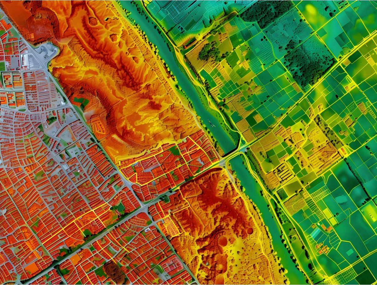
HSI used for multiple applications including for remote sensing © domi002 - stock.adobe.com

Spectroscopy and hyperspectral imaging (HSI) are powerful techniques that capture detailed spectral information across multiple wavelengths, enabling precise analysis of materials and environments. HSI extends traditional spectroscopy by combining spatially-resolved data with chemical and optical data, allowing for the identification of chemical composition with structural information, revealing subtle property variations across complex surfaces. HSI is widely applied in fields such as remote sensing, environmental analysis, medicine, pharmaceuticals, forensics, material science, agriculture, and food science, driving advancements in research, development, and quality control. The following articles on HSI have been published in Spectroscopy and the links to the original articles are given below.
Unlocking the Power of Hyperspectral Imaging: A Game-Changer for Agriculture, Medicine, and More
Hyperspectral imaging (HSI) is revolutionizing fields such as agriculture, food safety, and medical analysis by providing high-resolution spectral data. This emerging technology is proving invaluable in diverse applications, including plant stress detection, weed discrimination, and flood management. A new review explores HSI’s fundamental principles, applications, and future research directions. HIS is a cutting-edge technology that merges spectroscopy and imaging, is rapidly becoming a powerful tool in sectors ranging from agriculture to healthcare. By capturing spectral data for every pixel in an image, HSI creates a "hypercube" that offers unparalleled insights into the chemical and physical properties of materials. In a recent review published in Heliyon, researchers from various international institutions explored the transformative potential of HSI across multiple industries, emphasizing its value in areas such as food safety, medical analysis, water resource management, and more (1).
New Hyperspectral Imaging Database Enhances Human Skin Research
Researchers from the University of Minho (Portugal) have developed a hyperspectral imaging database of human facial skin, aimed at improving various scientific applications such as psychophysics-based research and material modeling. The database includes 29 participants with diverse skin tones, providing detailed spectral reflectance data under controlled conditions. A team from the Physics Center of Minho and Porto Universities (CF-UM-UP) at the University of Minho (Portugal), led by Andreia E. Gomes, Sérgio M. C. Nascimento, and João M. M. Linhares, has created a hyperspectral imaging database to capture the spectral characteristics of human facial skin. This comprehensive dataset is intended to support a variety of applications, from medical diagnostics to digital rendering, by providing precise, non-invasive measurements of skin reflectance. The research, published in Applied Spectroscopy, offers new insights into the spectral variations in human skin across different tones and regions of the face (2).
Using Hyperspectral Imaging to Analyze the Chemical Composition of Pet Food
In an effort to improve the quality of pet food, a recent research collaboration from Beijing, China, implemented a new approach to analyze the chemical composition of different types of pet food. The findings were published in Microchemical Journal (3). HSI was used to assess the chemical composition of pet food by capturing detailed spectral information across a wide range of wavelengths. This technology allows for the identification and quantification of various components, such as proteins, fats, carbohydrates, and moisture, without the need for physical contact or destructive sampling. By analyzing the specific spectral signatures of these components, HSI provides a comprehensive chemical profile of the pet food, ensuring quality control and consistency in formulation. This method can detect contaminants, adulterants, and nutritional imbalances, contributing to safer and healthier pet food products. The non-invasive nature of HSI also makes it a valuable tool for real-time monitoring during the manufacturing process, leading to enhanced efficiency and product quality (3).
Cutting-Edge vis-NIR Hyperspectral Imaging Enhances Bloodstain Identification in Forensic Science
Forensic scientists have made significant strides in bloodstain identification, leveraging advanced hyperspectral imaging and machine learning to distinguish between human and animal bloodstains with remarkable accuracy. In a pioneering study published in Applied Spectroscopy, researchers from the Academy of Criminal Investigation, Yunnan Police College, the Department of Forensic Science, Fujian Police College, and the Faculty of Science, Kunming University of Science and Technology have introduced a novel method for identifying bloodstains. The method, which combines HSI technology with the extreme learning machine (ELM) algorithm, has demonstrated superior performance in distinguishing bloodstains from different species, such as human, chicken, and pig blood (4).
Blood evidence plays a crucial role in solving criminal cases. However, identifying bloodstains at crime scenes can be challenging, especially when suspects attempt to destroy or obscure this vital evidence. Traditional forensic methods for blood identification rely on chemical and biochemical techniques that are often destructive, expensive, and time-consuming. This new approach aims to overcome these limitations by providing a rapid, non-destructive alternative (4).
Hyperspectral Imaging: An Examination of an Emerging Field in Spectroscopy
HSI is an advanced technique that captures and processes information across a wide range of the electromagnetic spectrum. Unlike traditional imaging methods that capture images in just three primary colors (red, green, and blue), HSI divides the spectrum into many more bands, often extending into the infrared and ultraviolet regions (5). This allows for the identification and analysis of materials and substances based on their spectral signatures, which are unique patterns of reflectance or emission at different wavelengths. Applications of HSI span various fields, including agriculture for crop monitoring, environmental science for detecting pollutants, medical diagnostics for identifying tissue abnormalities, and defense for surveillance and target identification. The technology's ability to provide detailed spectral and spatial information simultaneously makes it a powerful tool for enhancing the understanding and management of complex systems and processes (5).
Fruits’ Maturity Stages Measured Using Portable Hyperspectral Imagers
Scientists from Nanjing Forestry University recently used a portable hyperspectral imager to help detect maturity stages in Camellia oleifera fruits. Their findings were published in Spectrochimica Acta Part A: Molecular and Biomolecular Spectroscopy (6). Camellia oleifera fruits, which are extensively cultivated in various regions in southern China, are considered one of the four major woody oil crops globally, putting it alongside olives, palms, and coconuts. To obtain the highest amounts of seed yield and oil content, scientists must determine the optimal harvesting stage criteria for Camellia oleifera. This need has led to efforts by researchers to accurately identify the maturity stage of Camellia oleifera fruits.
Currently, this process is complex, and it is prone to being affected by different intrinsic and environmental factors. To rectify this problem, the scientists behind this study proposed a non-invasive detection method based around HSI technology. HSI integrates spectral and imaging techniques to simultaneously record spectral and spatial information, with the approach being used most often in the food industry for the nondestructive assessment of food quality. For example, a recent LCGC International piece focused on how hyperspectral imaging could be used to gather data on bruises in Fuji apples, predicting when these imperfections were caused during the delivery process and how much the bruises affect the overall quality of the apples (6). As a result, HSI is the best method for assessing the internal quality of fruit and its maturity stage while allowing for the acquisition of comprehensive sample information and generating large amounts of spectral and spatial data.
How Hyperspectral Imaging Can Evaluate Asian Soybean Rust Severity
Soybean rust, also known as Asian soybean rust, is an area of concern in agriculture applications. Asian soybean rust is caused by Phakopsora pachyrhizi (P. pachyrhizi), which is a pathogen that moves aggressively and has spread across several continents (7). Many agriculture yield losses can be directly attributed to this pathogen. In fact, yield losses have been reported in the United States to the tune of 10–80% since the pathogen was first detected in the United States in 2004. A new study published in Spectrochimica Acta Part A: Molecular and Biomolecular Spectroscopy explores this issue extensively. Led by Paulo Eduardo Teodoro and his team at the Federal University of Mato Grosso do Sul (UFMS), the research explored the potential of using machine learning (ML) and remote sensing to improve detection of Asian soybean rust (7). The research team conducted their experiment during the 2022–2023 harvest season.
Persimmon Leaves’ Contents Determined Using Hyperspectral Imaging
Using visible and near-infrared (Vis-NIR) HSI, scientists from Instituto Valenciano de Investigaciones Agrarias (IVIA) in Valencia, Spain were able to determine macro- and micronutrient contents rapidly and non-destructively in persimmon leaves. Their findings were published in Agriculture (8). Persimmon, a natively Chinese crop that mainly grows in tropical and subtropical areas, has grown exponentially in Spain in recent years. With its popularity comes a need for fair handling practices during growing season, including optimum nutrient management according to the trees’ nutritional statuses at each phenological stage. However, little research has been done on the nutritional requirements of persimmon. Macronutrients (nitrogen [N], phosphorus [P], potassium [K], to name a few) are major constituents of cell structures and organic compounds and are thusly needed in large amounts. In contrast, micronutrients (zinc [Zn], manganese [Mn], copper [Cu], as examples) are involved in the metabolic and enzymatic processes and are needed in small amounts. There has been little work to measure soil capacity for delivering these nutrients, which has pushed research towards proper crop monitoring.
In a new technology effort to tackle postharvest losses caused by invasive pests, researchers at the University of Kentucky, led by Alfadhl Y. Khaled, Nader Ekramirad, Kevin D. Donohue, et al., have unveiled a research study utilizing non-destructive HSI and machine learning to predict and manage the physicochemical quality attributes of apples during storage, specifically addressing the impact of codling moth infestation. The study, titled "Non-Destructive Hyperspectral Imaging and Machine Learning-Based Predictive Models for Physicochemical Quality Attributes of Apples during Storage as Affected by Codling Moth," was published in the journal Agriculture (9).
The research employed near-infrared HSI and machine learning models, utilizing partial least squares regression (PLSR) and support vector regression (SVR) methods. Data preprocessing involved Savitzky–Golay smoothing filters and standard normal variate (SNV), followed by outlier removal using the Monte Carlo sampling method. The study revealed significant effects of CM infestation on near-infrared (NIR) spectra, showcasing the potential impact of pests on apple quality (9).
The purpose of this work is to achieve rapid and nondestructive determination of tilapia fillets storage time associated with its freshness. Here, we investigated the potential of HSI combined with a convolutional neural network (CNN) in the visible and near-infrared region (vis-NIR or VNIR, 397−1003 nm) and the shortwave near-infrared region (SWNIR or SWIR, 935−1720 nm) for determining tilapia fillets freshness. Hyperspectral images of 70 tilapia fillets stored at 4 ℃ for 0–14 d were collected (10). Various machine learning algorithms were employed to verify the effectiveness of CNN, including partial least-squares discriminant analysis (PLS-DA), K-nearest neighbor (KNN), support vector machine (SVM), and extreme learning machine (ELM). Their performance was compared from spectral preprocessing and feature extraction. The results showed that PLS-DA, KNN, SVM, and ELM require appropriate preprocessing methods and feature extraction to improve their accuracy, while CNN without the requirement of these complex processes achieved higher accuracy than the other algorithms. CNN achieved accuracy of 100% in the test set of VNIR, and achieved 87.30% in the test set of SWIR, indicating that VNIR HSI is more suitable for detection freshness of tilapia. Overall, HSI combined with CNN could be used to rapidly and accurately evaluating tilapia fillets freshness (10).
After a brief introduction to the technique, this review will discuss the state-of-the-art NIR imaging instrumentation and its applications, including applications of an ordinary NIR imaging system to solvent diffusion into a polymer. Other imaging applications discussed include the use of a portable NIR imaging system in the pharmaceutical industry, high-speed and wide-area monitoring of polymers, and imaging for fish embryos development research. Also discussed is an imaging-type two-dimensional Fourier spectroscopy (ITFS) system and its application to medaka (Japanese rice fish, Oryzias latipes) egg development (11).
Using Hyperspectral Imaging in Food Analysis
In a recent study published in Spectrochimica Acta Part A: Molecular and Biomolecular Spectroscopy, researchers from the Beijing Technology and Business University used hyperspectral technology to analyze different types of wheat flour for quantification and quality (12). Wheat flour is an important ingredient in many food products around the world. Its quality and consistency are important because it ensures the quality of food products. The researchers combined several methods including hyperspectral technology, advanced data analysis methods, and machine learning to distinguish between five different types of wheat flour.
In this study, the researchers use hyperspectral imaging, which captures information across a broad spectrum of wavelengths. The research team established an analysis model based on the reflectance of wheat flour samples at wavelengths spanning from 968 nm to 2576 nm (12).
Identifying Freshness of Shrimp Following Refrigeration Using Near-Infrared Hyperspectral Imaging
Shrimp tends to deteriorate during the refrigeration process. To monitor the freshness of shrimp during refrigeration, near-infrared (NIR) HSI was utilized to non-destructively identify the freshness of shrimp. In the process, three preprocessing methods (multivariate scatter correction [MSC], standard normal variate [SNV], and direct orthogonal signal correction [DOSC]) were employed to preprocess the full-wavelength spectral data, and three characteristic wavelength extraction algorithms (competitive adaptive reweighted sampling [CARS], and random forest [RF] simulated annealing [SA]) were used to extract the best-pre-processed data. Because extreme learning machine (ELM) and kernel extreme learning machine (KELM) are easily affected by parameters, ELM (based on teaching-learning-based optimization [TLBO]) and KELM (based on teaching-learning-based optimization [TLBO]) were proposed. In this study, four discriminant models (ELM, TLBO– ELM, KELM, and TLBO–KELM) were used for the full wavelength modeling analysis and the characteristic wavelength modeling analysis. In this work, the results of the final selected models are presented (13).
References
(1) Workman, Jr., J. Unlocking the Power of Hyperspectral Imaging: A Game-Changer for Agriculture, Medicine, and More. Spectroscopyonline.com. Available at: https://www.spectroscopyonline.com/view/unlocking-the-power-of-hyperspectral-imaging-a-game-changer-for-agriculture-medicine-and-more (accessed 2024-12-02).
(2) Workman, Jr., J. New Hyperspectral Imaging Database Enhances Human Skin Research. Spectroscopyonline.com. Available at: https://www.spectroscopyonline.com/view/new-hyperspectral-imaging-database-enhances-human-skin-research (accessed 2024-12-02).
(3) Wetzel, W. Using Hyperspectral Imaging to Analyze the Chemical Composition of Pet Food. Spectroscopyonline.com. Available at: https://www.spectroscopyonline.com/view/using-hyperspectral-imaging-to-analyze-the-chemical-composition-of-pet-food (accessed 2024-12-02).
(4) Workman, Jr., J. Cutting-Edge vis-NIR Hyperspectral Imaging Enhances Bloodstain Identification in Forensic Science. Spectroscopyonline.com. Available at: https://www.spectroscopyonline.com/view/cutting-edge-vis-nir-hyperspectral-imaging-enhances-bloodstain-identification-in-forensic-science (accessed 2024-12-02).
(5) Wetzel, W. Hyperspectral Imaging: An Examination of an Emerging Field in Spectroscopy. Spectroscopyonline.com. Available at: https://www.spectroscopyonline.com/view/hyperspectral-imaging-an-examination-of-an-emerging-field-in-spectroscopy (accessed 2024-12-02).
(6) Acevedo, A. Fruits’ Maturity Stages Measured Using Portable Hyperspectral Imagers. Spectroscopyonline.com. Available at: https://www.spectroscopyonline.com/view/fruits-maturity-stages-measured-using-portable-hyperspectral-imagers (accessed 2024-12-02).
(7) Wetzel, W. How Hyperspectral Imaging Can Evaluate Asian Soybean Rust Severity. Spectroscopyonline.com. Available at: https://www.spectroscopyonline.com/view/how-hyperspectral-imaging-can-evaluate-asian-soybean-rust-severity (accessed 2024-12-02).
(8) Acevedo, A. Persimmon Leaves’ Contents Determined Using Hyperspectral Imaging. Spectroscopyonline.com. Available at: https://www.spectroscopyonline.com/view/persimmon-leaves-contents-determined-using-hyperspectral-imaging (accessed 2024-12-02).
(9) Spectroscopy Staff. Cutting-Edge Technology Safeguards Apple Quality: Hyperspectral Imaging and Machine Learning to Combat Codling Moth Infestation. Spectroscopyonline.com. Available at: https://www.spectroscopyonline.com/view/cutting-edge-technology-safeguards-apple-quality-hyperspectral-imaging-and-machine-learning-to-combat-codling-moth-infestation (accessed 2024-12-02).
(10) Tang, S.; Li, P. Hyperspectral Imaging Combined with Convolutional Neural Network for Rapid and Accurate Evaluation of Tilapia Fillet Freshness. Spectroscopyonline.com. Available at: https://www.spectroscopyonline.com/view/hyperspectral-imaging-combined-with-convolutional-neural-network-for-rapid-and-accurate-evaluation-of-tilapia-fillet-freshness (accessed 2024-12-02).
(11) Ozaki, Y.; Ishigaki, M. Frontiers of NIR Imaging. Spectroscopyonline.com. Available at: https://www.spectroscopyonline.com/view/frontiers-of-nir-imaging (accessed 2024-12-02).
(12) Spectroscopy Staff. Using Hyperspectral Imaging in Food Analysis. Spectroscopyonline.com. Available at: https://www.spectroscopyonline.com/view/using-hyperspectral-imaging-in-food-analysis (accessed 2024-12-02).
(13) Ye, R.; Liu, C. Identifying Freshness of Shrimp Following Refrigeration Using Near-Infrared Hyperspectral Imaging. Spectroscopyonline.com. Available at: https://www.spectroscopyonline.com/view/identifying-freshness-of-shrimp-following-refrigeration-using-near-infrared-hyperspectral-imaging (accessed 2024-12-02).
NIR Spectroscopy Explored as Sustainable Approach to Detecting Bovine Mastitis
April 23rd 2025A new study published in Applied Food Research demonstrates that near-infrared spectroscopy (NIRS) can effectively detect subclinical bovine mastitis in milk, offering a fast, non-invasive method to guide targeted antibiotic treatment and support sustainable dairy practices.
Karl Norris: A Pioneer in Optical Measurements and Near-Infrared Spectroscopy, Part II
April 21st 2025In this two-part "Icons of Spectroscopy" column, executive editor Jerome Workman Jr. details how Karl H. Norris has impacted the analysis of food, agricultural products, and pharmaceuticals over six decades. His pioneering work in optical analysis methods including his development and refinement of near-infrared spectroscopy, has transformed analysis technology. In this Part II article of a two-part series, we summarize Norris’ foundational publications in NIR, his patents, achievements, and legacy.
AI-Powered SERS Spectroscopy Breakthrough Boosts Safety of Medicinal Food Products
April 16th 2025A new deep learning-enhanced spectroscopic platform—SERSome—developed by researchers in China and Finland, identifies medicinal and edible homologs (MEHs) with 98% accuracy. This innovation could revolutionize safety and quality control in the growing MEH market.