Thermally-Induced Structural Changes in Proteins Measured by ATR Spectroscopy
Infrared ATR spectroscopy has recently gained recognition as a viable technique for protein structural analysis due to its high information content, rapid sampling rate, and minimal sample preparation.
Infrared ATR spectroscopy has recently gained recognition as a viable technique for protein structural analysis due to its high information content, rapid sampling rate, and minimal sample preparation. Much focus has been placed on studying protein unfolding, where infrared allows analysis of secondary structural changes on a timescale unachievable by other methods.This note explores the use of a multiple reflection ATR to analyze small amounts of a protein undergoing secondary structural changes in response to thermal perturbation.
Experimental
HarrickConcentratIR2™ (see Figure 1) with its Si ATR crystal and heated liquid cell was installed and aligned in a commercial FTIR spectrometer with an MCT detector. Spectra were collected at 4 cm-1 resolution and signal averaged over 64 scans. The sample, 750 μg/mL BGG in a 0.9% sodium chloride solution with sodium azide, was injected into the liquid cell after it had stabilized ata temperature of 100 °C. Immediately after injection, the cell was capped to prevent evaporation and an initial spectrum was collected. Additional spectra were collected at timed intervals.

Figure 1: ConcentratIR2⢠Multiple Reflection ATR with its Heated Flow Cell and Temperature Controller.
Results and Discussion
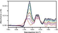
Figure 2 shows the spectral changes in the amide I band in the 1600–1700 cm-1 region. The most dramatic effect is a decrease in the intensity of the band around 1670 cm-1, which occurs fairly rapidly after heating, and an increase in the intensity of the band at 1620 cm-1.

Figure 2: The ATR spectra of BGG at 100 °C at 0 (lowest peak),1, 2, 3, 4, 5, 6, 7, 8, 9, 20, and 30 (highest peak) min after injection of sample into cell.
The amide I band consists of overlapping components that reflect secondary structures due to the hydrogen-bonded backbone of the protein. The most common secondary structures are the α-helices, which have a characteristic band around 1660 cm-1 and the β sheets, which typically show bands around 1620–1640 cm-1. So the observed spectral changes are consistent with the relatively quick thermal degradation of the weaker α-helices followed by the slower formation of β sheets.
Conclusion
The ConcentratIR2™, a multiple reflection ATR, can be used effectively to examine thermally-induced secondary structural changes in proteins. Since the active area of the ATR element is only 4-mm in diameter, only a minute quantity of the sample is required for analysis, making the ConcentratIR2™ extremely useful in situations where only limited quantities of sample are available.

Harrick Scientific Products
Box 277, Pleasantville, NY 10570
tel. (800) 248-3847, fax (914) 747-7209
Website: www.harricksci.com

A Seamless Trace Elemental Analysis Prescription for Quality Pharmaceuticals
March 31st 2025Quality assurance and quality control (QA/QC) are essential in pharmaceutical manufacturing to ensure compliance with standards like United States Pharmacopoeia <232> and ICH Q3D, as well as FDA regulations. Reliable and user-friendly testing solutions help QA/QC labs deliver precise trace elemental analyses while meeting throughput demands and data security requirements.