Analysis of Transdermal Patch Layers via Confocal Raman MicroscopyAnalysis of Transdermal Patch Layers via Confocal Raman Microscopy
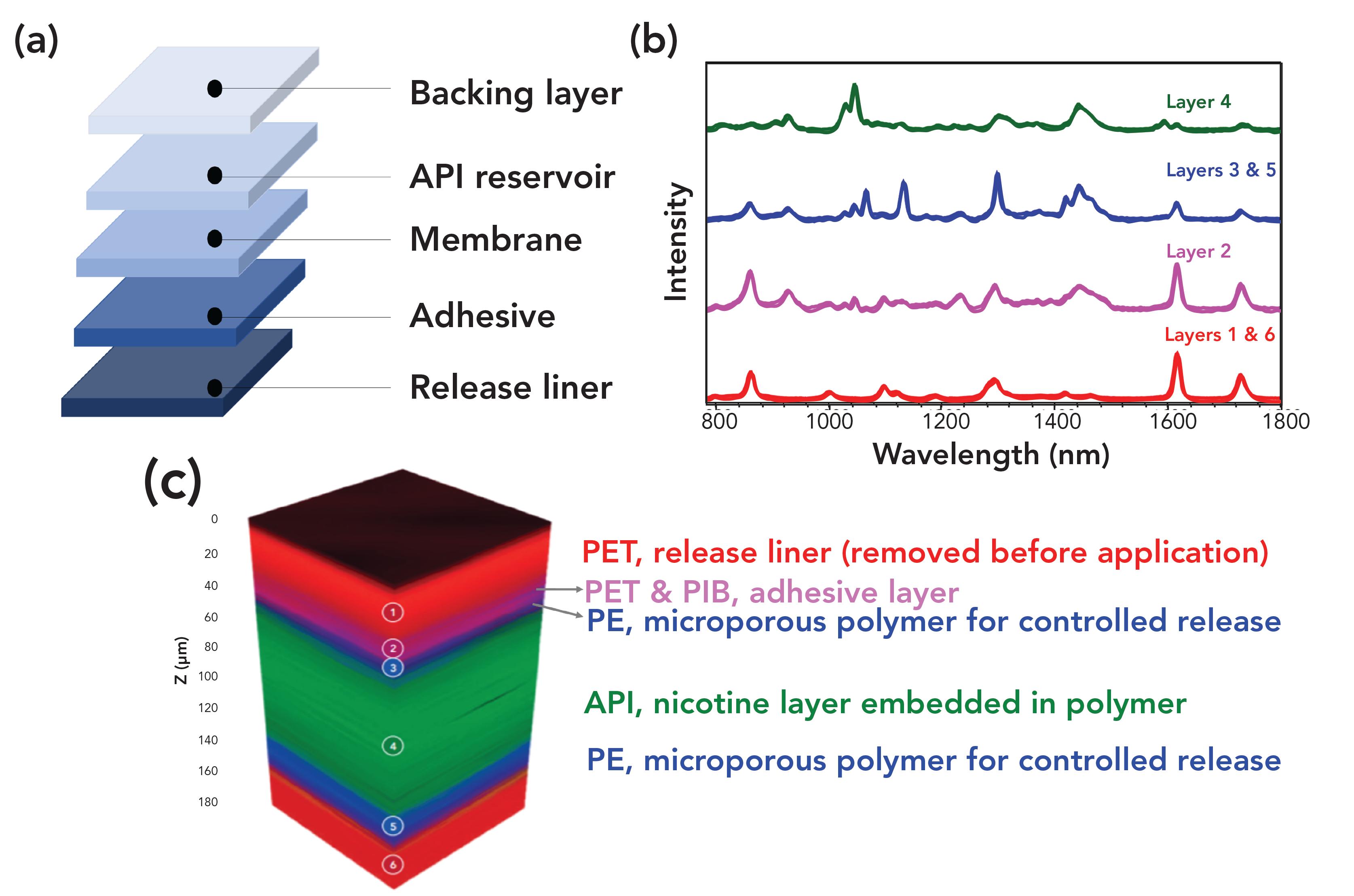
A transdermal patch is a method of drug delivery through the skin into the bloodstream. The patch, shown in Figure 1(a), contains a drug that absorbs into the skin when attached, releasing a consistent and controlled amount of medication into the body. One of the most common uses for transdermal patches is to help combat nicotine addiction. Nicotine replacement therapy releases small amounts of nicotine throughout the day, without the additional harmful components of cigarettes.
In this application note, we study a nicotine patch to reveal the active pharmaceutical ingredient (API) as well as the polymer layers encapsulating it via confocal Raman microscopy.
Experimental conditions
An RMS1000 Raman microscope, equipped with a 785-nm laser, was used to measure a 3D Raman map of the transdermal nicotine patch. The nicotine patch was purchased from a pharmacy. The RMS1000 can acquire 3D Raman maps thanks to its confocal pinhole. The pinhole acts as a spatial filter in the axial (Z) direction by blocking the Raman scatter from above and below the focal plane and is essential for 3D mapping. Without the pinhole, there is no axial spatial filtering, and Raman scatter from the entire axial volume of the sample would be collected with no axial resolution.
Figure 1: a) Layers of a transdermal patch, b) spectra from each layer of the patch, c) 3D render of the Raman map with each layer identified.

Results
The transdermal patch analyzed contained nicotine as the active ingredient surrounded by polymers. By carrying out a Raman 3D map we can visualize the different material layers present in patch, Figure 1(b). The 3D Raman map, shown in Figure 1(c), reveals that the patch contains 6 layers; the spectrum of each layer can be extracted and analyzed for identification. The outer layers of the patch, layers 1 and 6, shown in red, can be identified as poly(ethylene terephthalate) (PET) using the KnowItAll Raman Database. The 3D map is colored based on peaks specific to the material identified; for example for the layers of PET the peak at 1293 cm-1 was selected.
Layer 2 can be identified as PET/polyisobutylene, and layers 3 and 5, which surround the API, were determined to be polyethylene (PE). Finally, in the middle of the polymers is a thick layer of the API, nicotine, embedded in polymer reservoir (ethylene).
Conclusions
This application note demonstrated how the RMS1000 Raman microscope can be used for 3D mapping of a transdermal patch. The API, in this case nicotine, can be visualized as can the surrounding polymers responsible for protecting and ensuring API release. In this way confocal Raman microscopy is a useful technique for pharmaceutical laboratories to conduct quality control on their products, ensuring they have been produced correctly without contaminants, and help monitor the stability of adhesive layers.
Edinburgh Instruments
2 Bain Square, Kirkton Campus, EH54 7DQ, UK
Tel: +44 (0) 1506 425 300 Fax: +44 (0) 1506 425 320
www.edinst.com


Hole Extraction in Perovskite Solar Cells
December 16th 2024Efficient charge extraction is crucial for high-efficiency solar cells. Electron and hole extraction layers optimize cell performance. PL spectroscopy, proportional to carrier number, is ideal for comparing extraction layer efficiency.