Analysis of Polymorphic Materials on the RISE
RISE microscopy, the combination of Raman imaging and scanning electron microscopy, is a powerful new analytical method for the analysis and interrelation of the structure and composition of microscopic samples. The integration of both imaging techniques in one instrument avoids shuttling the sample from one microscope to another. Here we demonstrated its usefulness in the identification, discrimination, and localization of polymorphs of titanium dioxide.
RISE microscopy is a novel correlative microscopy technique that combines confocal Raman imaging and scanning electron microscopy. We have developed an approach to fuse these methods into one instrument, hence avoiding the inevitable complication of sample transfer and re-identification of the investigated area when using two separate instruments. With the example of titanium(IV)-dioxide (TiO2) we show that RISE microscopy can easily and reliably distinguish and visualize polymorphic modifications of materials.
Linking Molecular Composition to Structure
Compound materials that can occur in more than one variety of crystal structure are called polymorphs. Structural polymorphism plays an important role not only in materials research and development but also in the production of pharmaceutical, agrochemical, and pigment substances because the manner of atomic bonding greatly influences the properties of a material. Techniques such as AFM, NMR, and IR-spectroscopy are employed to distinguish polymorphs. While high resolution imaging methods can visualize the morphology of a sample, spectroscopic approaches enable the identification of the chemical substances therein. Correlating both with RISE microscopy, the sample's structural features can be directly linked to its molecular composition.
Titanium dioxide is studied intensively due to its interesting chemical and optical properties. It is widely employed in photocatalysis, electrochemistry, and chemical catalysis while also being the most important pigment in coatings, paints, cosmetics, and as E 171 in food products. TiO2 occurs in eight modifications, with the most commonly used being anatase and rutile. Depending on the application, one crystalline form or the other can gain in importance. Hence clear discrimination of TiO2 polymorphs is an important issue in the products' manufacturing process and usage.
For RISE microscopy anatase and rutile powders were mixed 1:1, ground, dissolved in water, and measured. Raman imaging was performed with a WITec alpha300 microscope, for scanning electron microscopy a Tescan SEM from Brno, Czech Republic was used. Both microscopes were integrated into one instrument. In this hybrid system the sample remains within the vacuum chamber and is automatically and remotely transferred from one measurement position to the other. With this combination, the overlaying of SEM and confocal Raman images is easy, precise, and straightforward.
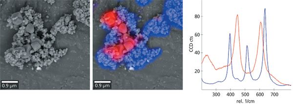
Both modifications of TiO2 are of the same elemental composition, which can be detected by the energy dispersive X-ray (EDX) functionality incorporated with the SEM (data not shown). However, EDX can't offer insight regarding the arrangement and coordination of the detected atoms and their chemical bonds. That particular characterization is best accomplished with Raman spectroscopy. Here, anatase and rutile were easily distinguished from one another by their Raman spectra at wavenumbers between 300 and 800 rel. 1/cm (Figure 1c). Scanning electron microscopy revealed TiO2 particles of various sizes (Figure 1a). By correlating this information with the Raman spectra generated at each pixel of the image it was possible to assign rutile to the larger and anatase to the smaller particles (Figure 1c). To our knowledge this is the first direct imaging of the spatial distribution of anatase and rutile phases of TiO2 in a mixture of both.

Figure 1: Two modifications of TiO2, anatase and rutile, were imaged with a SEM (a) and a confocal Raman microscope. Both images were then overlaid to yield the RISE image (b). In the Raman spectrum (c) anatase (blue) can be easily distinguished from rutile (red). Image parameters: 12 µm × 12 µm, 150 × 150 pixels = 22,500 spectra, integration time 0.037 s per spectrum.
Conclusion
RISE microscopy, the combination of Raman imaging and scanning electron microscopy, is a powerful new analytical method for the analysis and interrelation of the structure and composition of microscopic samples. The integration of both imaging techniques in one instrument avoids shuttling the sample from one microscope to another. Here we demonstrated its usefulness in the identification, discrimination, and localization of polymorphs of titanium dioxide.

WITec GmbH
Lise-Meitner-Stra e 6, 89081 Ulm, Germany
tel. +49 (0) 731 149 700, fax +49 (0) 731 149 70 200
Website: www.witec.de
