Analysis of a Mixture Solution Using Silver Nanoparticles Based on Surface-Enhanced Raman Spectroscopy (SERS)
Scopolamine and promethazine can be used as a substitute for heroin. It is relatively fast and simple to use surface-enhanced Raman spectroscopy (SERS) for the detection and monitoring of drug usage. Silver nanosol is an enhanced substrate that is commonly used in the SERS technique; the Raman signal of the sample is amplified by the substrate. However, for the detection of mixtures, the competitive adsorption of different components relative to the substrate will result in difficult qualitative identification of spectra. By taking advantage of this defect, the adsorbed components of a mixture were separated from silver nanoparticles by centrifugation, thus changing the mixing ratio of the two components in the mixture. As a result, the proportion of the components with strong adsorption in the mixture is relatively reduced. Thus, the components with weak adsorption can be displayed in the spectrum of the mixed solution.
Heroin is one of the most widely used narcotics in the world, and it is a key target to be monitored because of its addictive quality and the difficulty in quitting the addiction (1–6). After long-term use, heroin will cause serious damage to the nervous system of the human body, and will also adversely affect judgment, possibly causing harm to the addict or society (4,7,8). Some drug rehabilitation institutions use less harmful drug-like substances as substitutes to reduce the suffering of drug addicts and damage to their nervous system (9–14). For example, a mixture of buprenorphine, scopolamine, and promethazine, referred to as BSP, has been used because its toxicity is much lower than that of heroin (15). However, long-term use of BSP will still cause great damage to the human body (9,10). Because the price of BSP is low and it is easily accessible, it has become an alternative drug for some drug users. Therefore, it is necessary to engage in the detection and monitoring of this new type of drug abuse.
Surface-enhanced Raman spectroscopy (SERS) is a combination of Raman spectroscopy and nanotechnology, and it can enhance the Raman signal by 7–10 orders of magnitude when the tested sample is near or adsorbed on a substrate at the nanometer scale (16–19). The Raman spectrum of each sample is specific, and can be qualitatively identified as the fingerprint of the sample.
There are two popular viewpoints about the enhancement mechanism of SERS: One viewpoint is electromagnetic enhancement, and the other viewpoint is chemical enhancement (17,20). Electromagnetic enhancement generally refers to the enhancement of Raman signals by the strong electromagnetic field generated by the local surface plasmon (LSPR) excited by the rough surface of metal materials or the surface of metal nanoparticles (21). The chemical mechanism refers to the contribution of Raman scattering independent of the electromagnetic environment (such as plasma polaritons). The increase in SERS signal is usually attributed to electron transfer between the adsorbed molecule and the substrate. Current research results show that the two enhancement mechanisms generally play a role at the same time, and that signal amplification effect of electromagnetic enhancement is greater than that of chemical enhancement. Based on these enhancement phenomena, the SERS signal can be improved to some extent through substrate design or gap control (22–25).
Surface-enhanced spectroscopy as a drug detection technology has become increasingly mature (26–29). Because of its characteristics, it is more suitable for the detection of drugs in solution than many other analytical techniques. Compared with other detection technologies, the advantages of SERS lie in the simple operation of the instrument, the uncomplicated pretreatment of the sample before detection, the fast detection time, and the convenience of the portable instrument for the in situ detection of samples (30).
For SERS detection of mixtures, there are two problems associated with direct detection: One is the overlapping of peaks, and the other is competitive adsorption (31). Overlap of spectral peaks is most likely to occur when the molecular configurations of the components of the mixture are relatively similar.
There are some differences in the molecular configuration of the two components we tested. They were distinguished from the spectrum of the mixture as described in our previous studies (15). From the spectral observation, there is no chemical reaction of the two components after mixing; that is, no new substance is formed. The current study focuses on eliminating the interference of competitive adsorption in practical detection.
Experimental
Apparatus
A BWS415-785H portable Raman spectrometer (B&W Tek, Inc.) was used to obtain the spectral data. The maximum working power of the Raman spectrometer was 0.3 W. The laser wavelength was 785 nm. After spectrum acquisition, the spectrum is smoothed and the baseline was corrected by the instrument software.
The morphology of nanoparticles was characterized by a high-resolution field emission scanning electron microscope (Hitachi Company). The dynamic light scattering test was measured by a PALS/90P particle size analyzer (Brookhaven Instruments Co., Ltd.). A Cary 4000 UV-vis spectrophotometer was also used (Agilent Technology Co., Ltd.).
Reagents
Potassium iodide (KI), silver nitrate (AgNO3), sodium citrate (C7H5Na3O7), and ascorbic acid (C6H8O6) were purchased from Sinophram Chemical Reagent Co., Ltd. Scopolamine hydrobromide powder (1G) and promethazine hydrochloride powder (5G) were purchased from Dalian Meilun Biotechnology Co., Ltd.
Sample and Substrates Preparation
The solutions of scopolamine and promethazine were obtained by diluting scopolamine hydrobromide and promethazine hydrochloride, respectively, with deionized water, are used to obtain solutions with different concentrations. Each solution is mixed with the substrate at the same time.
The preparation of the silver nano-substrate is as follows: Soluble silver salt nitrate (AgNO3) was used as an oxidant, and ascorbic acid was used as the reducing agent to prevent the aggregation of silver nanoparticles. Sodium citrate was used as the stable solution. Their concentrations in the initial solution were 0.168 mg/mL, 0.105 mg/mL, and 0.873 mg/mL. First, the water bath condition was prepared at 30 oC. Then, the three concentrations (soluble silver salt nitrate, ascorbic acid, and sodium citrate) were placed in the bath proportional to each other, then the solutions (sols) were stirred continuously for 15 min with a magnetic mixture and both sol and nanosol are used. Then, the temperature of the water bath was continually raised to the boiling state and stayed at that temperature level for 2 h until the color turned grayish-green. Then, the sol was refrigerated after cooling. Before using it, the sol needed to be centrifuged and purified to remove the smaller particles and improve the monodispersibility of the particles. Ultrasonic oscillation dispersion was performed before the test was used.
Results and Discussion
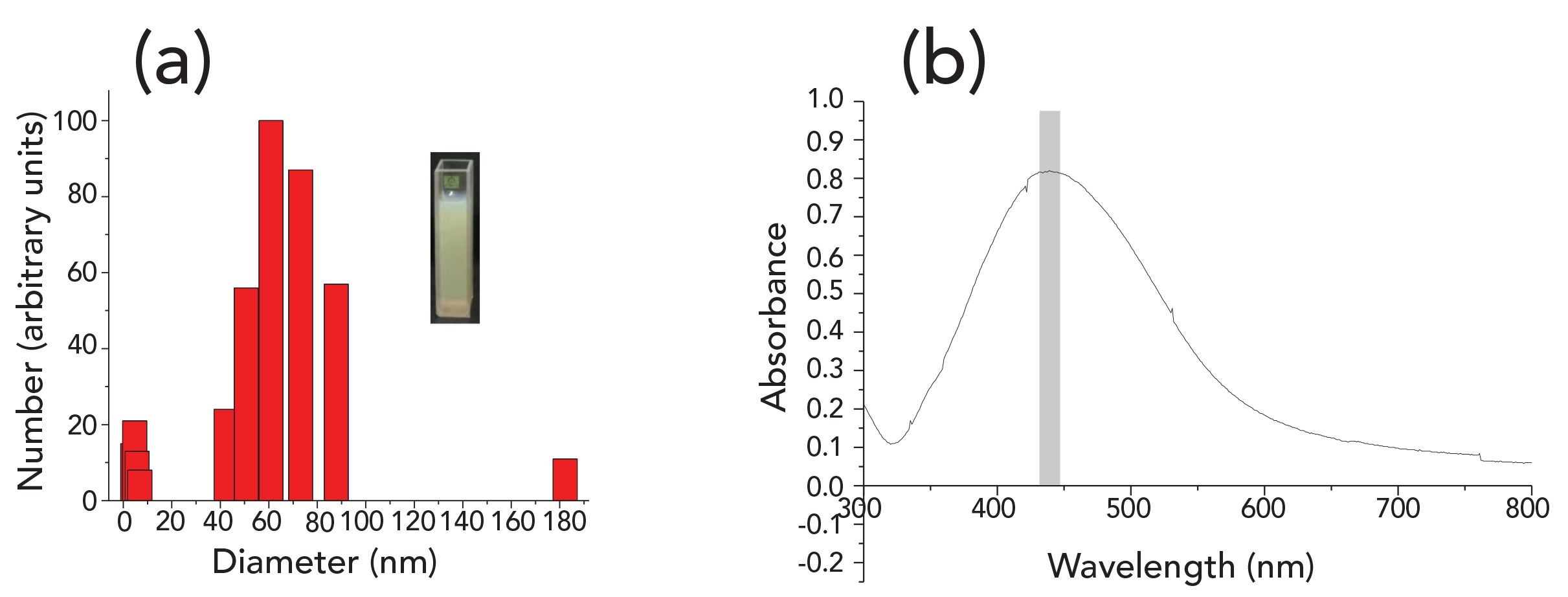
Figure 1 shows the SEM characterization of the silver sol substrate. The main process used is described in the literature (32). The silver nanoparticles are basically in a spherical state, and there is no mixing of other shapes of the substrate. Figure 2a shows the UV absorption spectrum, and the scanning range is from 300 nm to 800 nm. The absorption resonance peak is approximately 439 nm, which is consistent with the absorption peak of the silver sol. Figure 2b shows the statistical weight distribution of the particle size of the silver sol measured by dynamic light scattering (DLS) technology. Among them, 60 nm particles account for the largest proportion in the system, with its statistical weight being the total number of particles set to 100% and the statistical weight of other particle sizes compared with its value as the ordinate value of other particle sizes. It can be seen that several main particle sizes are distributed at 50–80 nm.
FIGURE 1: SEM image of silver sol substrate.
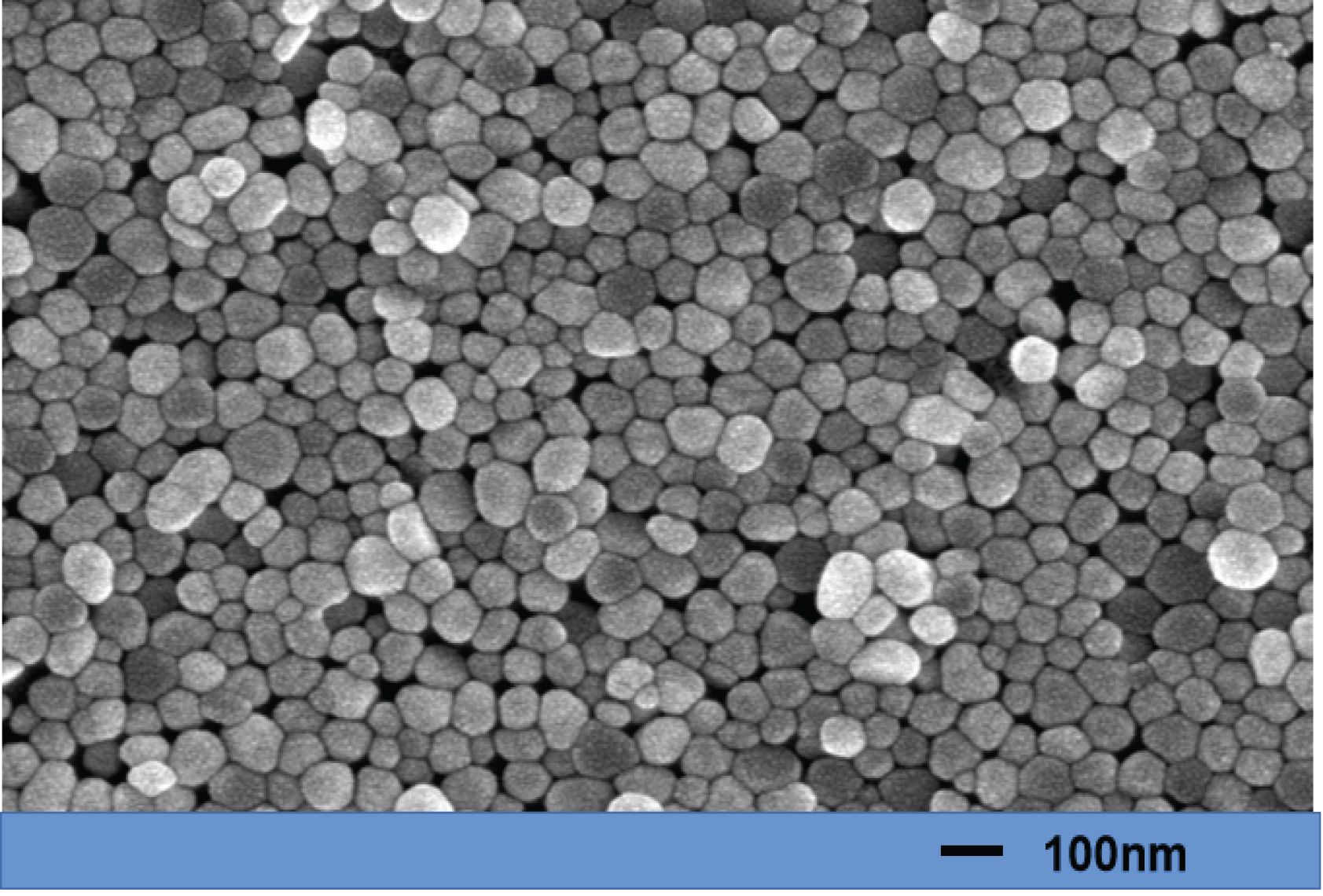
FIGURE 2: (a) Particle size distribution of silver sol; (b) UV-vis extinction spectra of silver sol.
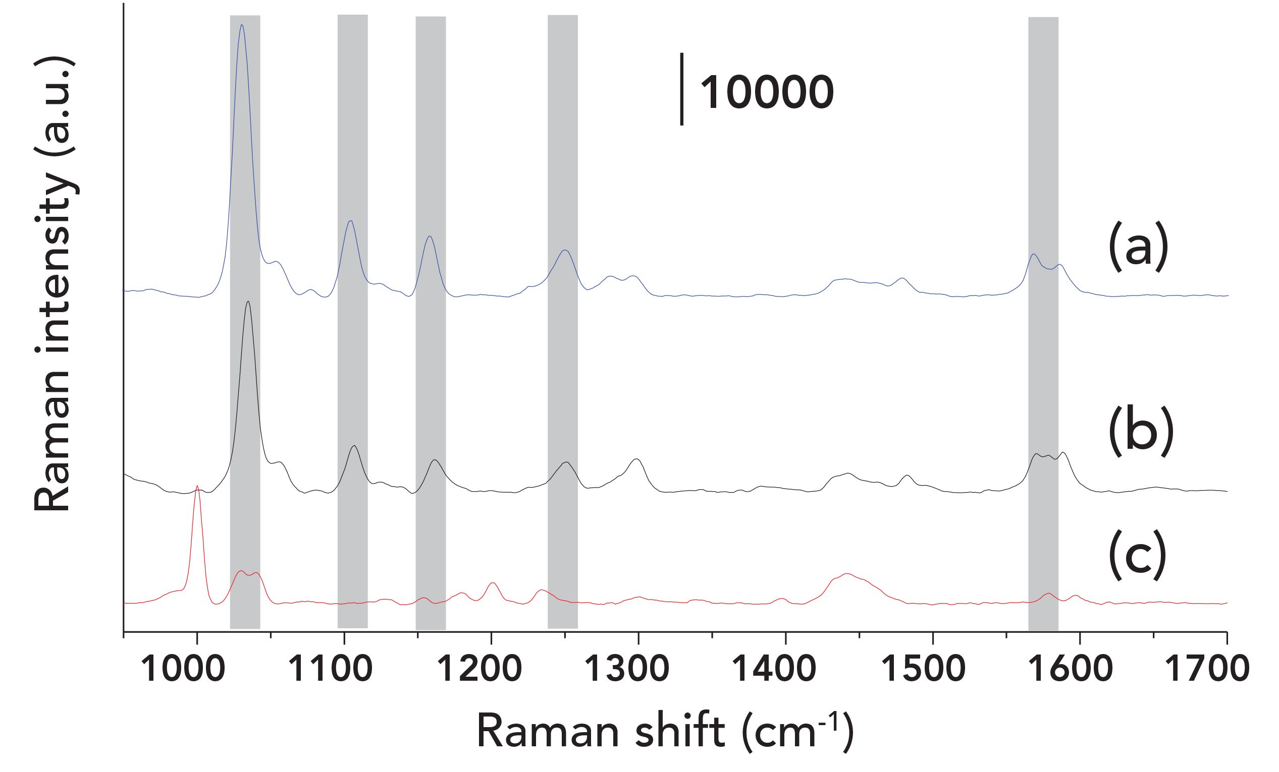
The SERS spectra of 10 ppm (3.5 x 10⁻⁵ M) promethazine and 6 ppm (2 x 10⁻⁵ M) scopolamine were used to compare the mixture, as shown in Figures 3a and 3c. It should be noted that because the signal of scopolamine is weak, the intensity of scopolamine is exaggerated by about three times. When the molar concentration ratio of the two mixtures was 1:1 (1.5 x 10⁻⁵ M), a weak Raman signal of scopolamine was detected at 1002 cm-1. As shown in Figure 3b, the characteristic peaks in the mixture mainly belong to promethazine. However, in the previous study, when the concentration of the two components was the same, the spectrum was mainly the spectrum of scopolamine (15). The difference is that in our current study, the mixture was in contact with the silver nanoparticles at the same time; in the previous study, scopolamine was first added to the silver nanosol (nanosolution), and then the promethazine was subsequently added. From the trend of these two spectra, we can deduce that there is a competitive adsorption phenomenon when they are mixed simultaneously. Promethazine will mask the characteristic peak of scopolamine at the same concentration, which is not conducive to identifying the spectrum of the mixture. What is more troublesome is that in the actual mixing ratio, the concentration of scopolamine is much lower than that of promethazine. To enhance the signal of scopolamine, we added potassium iodide into the mixture system to increase the strength of scopolamine.
FIGURE 3: SERS spectra of (a) promethazine 10 ppm (3.5 x 10⁻⁵ M); (b) mixture at concentration ratio of 1:1 (1.5 x 10⁻⁵ M); (c) scopolamine 6 ppm (2 x 10⁻⁵ M).
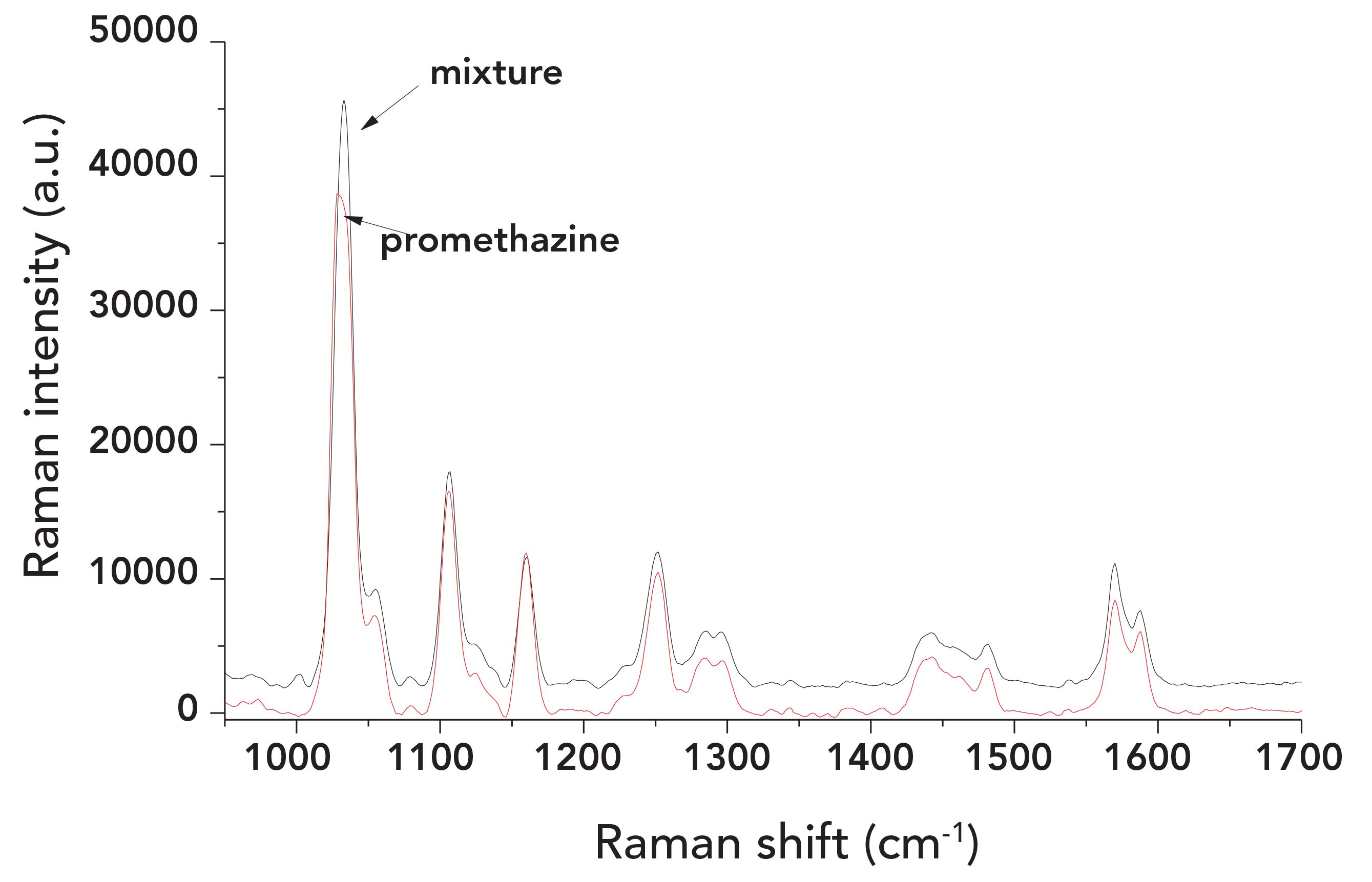
To improve the signal of scopolamine in the mixed solution in Figure 3, we added potassium iodide (1 M). As shown in Figure 4, the signal of scopolamine at 1002 cm-1 increases with the addition of electrolytes, but it greatly decreases compared with the detection of scopolamine at the same concentration alone. Moreover, the concentration ratio of the silver sol we used is further increased, and the signal should be stronger in theory than what is shown in Figure 4. The characteristic peaks of promethazine in the spectrum of the mixed solution are basically all displayed. Promethazine of the same concentration was added to the electrolytes for independent detection and is compared with the mixture spectrum, as shown in Figure 5. The position and intensity of the peak of the mixture spectrum are almost the same as that of promethazine alone. Table I compares the intensity of the mixture spectrum with that of promethazine when it is separately detected in several main characteristic peaks. The results show that the signal of promethazine was dominant even though both of them were enhanced. This further shows that the enhanced signal of the two is mainly caused by the molecules adsorbing on the silver nanoparticles. The addition of electrolytes did not change the adsorption ratio of the two but further reduced the distance between nanoparticles and enhanced their signal.
FIGURE 4: SERS spectra of mixture at concentration ratio of 1:1 (1.5 x 10⁻⁵ M) with potassium iodide (KI).
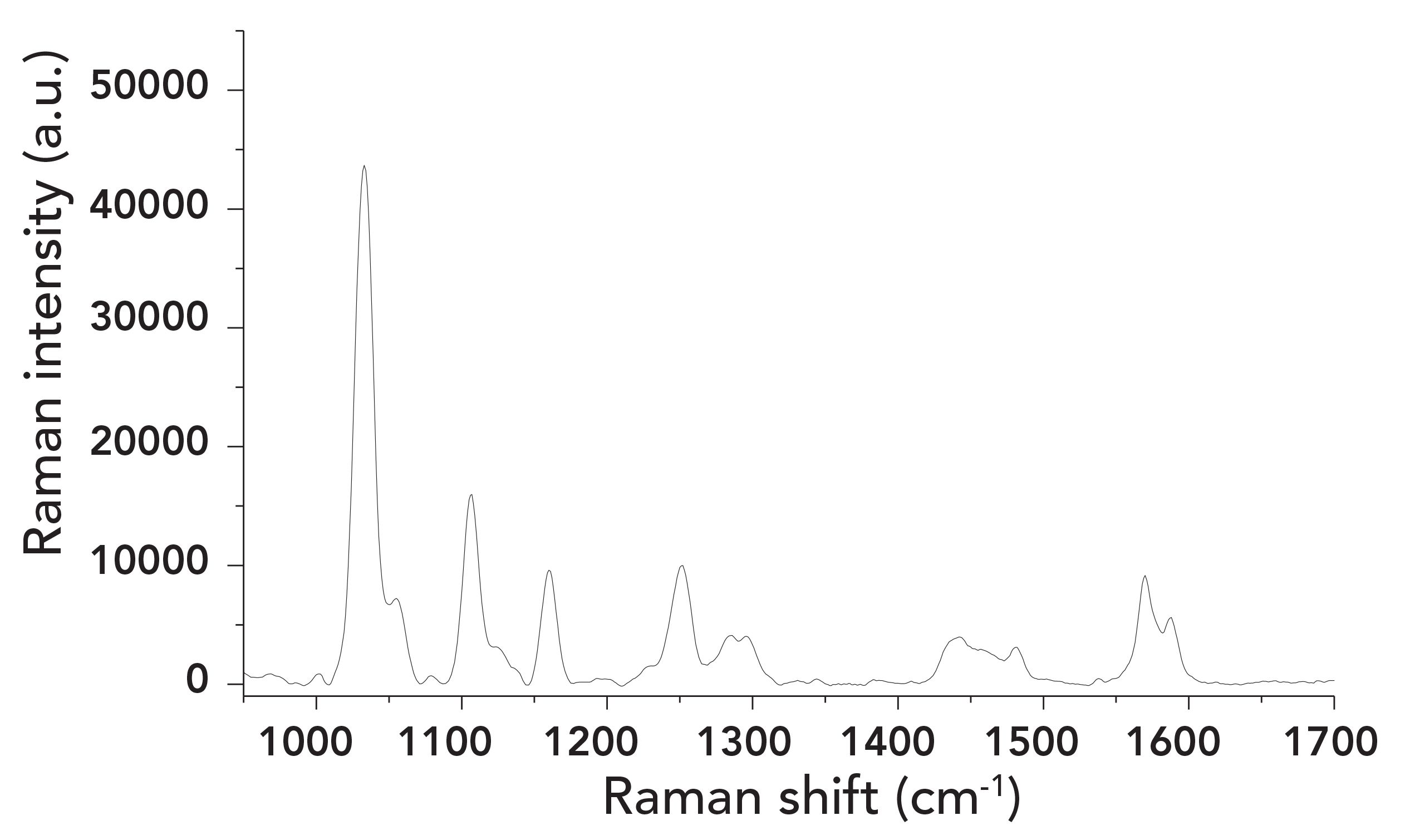
FIGURE 5: Comparison of SERS spectra between mixture and promethazine.

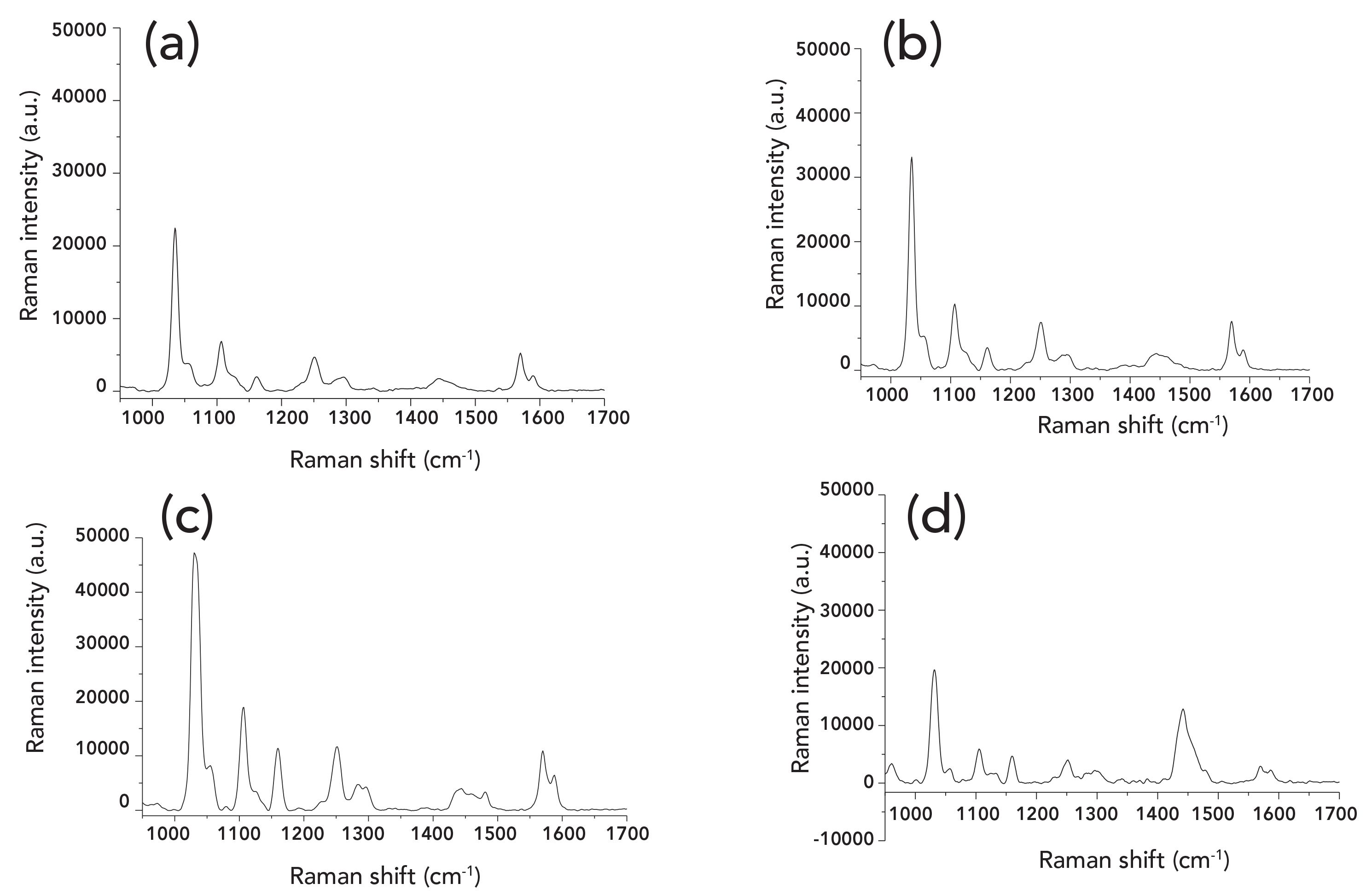
In the mixture with the same concentration, the signal of scopolamine is weak, which is not conducive to spectral recognition. The more difficult problem is that the concentration of promethazine with strong adsorption is much higher than that of scopolamine in the actual ratio. It can predict the difficulty of actual detection. To verify this assumption, we mixed them according to the actual ratio, in which the mass concentration of scopolamine is 10 ppm (3.3 x 10⁻⁵ M), and the doping of promethazine is 1670 ppm (5.9 x 10⁻³ M). Through SERS detection, as shown in Figure 6a, scopolamine in the actual ratio cannot be identified in the Raman spectrum. Only the characteristic peaks of promethazine can be identified, which is consistent with the expected results.
FIGURE 6: SERS spectra of mixture (a) original solution (promethazine: 5.9 ́ 10-3 M; scopolamine: 3.3 ́ 10-5 M) (b) the first centrifugation, (c) the second centrifugation, and (d) the last centrifugation.
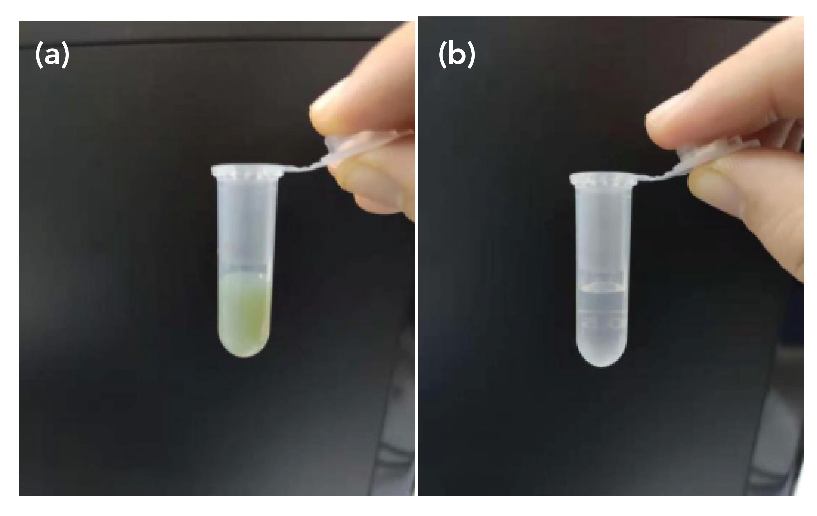
According to the experimental verification, the nanoparticles in the silver sol are relatively easy to separate from the solution, as shown in Figure 8. For example, most of the nanoparticles will be located at the bottom of the tube after high-speed centrifugation. Figure 8a shows the original sol solution, and the supernatant after centrifugation is shown in Figure 8b. We used this method in combination with the “defects” of competitive adsorption to separate the components of the mixture in solution. After the mixed solution was added to the silver sol and then centrifuged, the silver sol traveled to the bottom of the centrifuge tube. Some samples adsorbed on the silver nanoparticles were separated from the mixed solution.
FIGURE 8: (a) Silver sol before centrifugation, and (b) silver sol after centrifugation.
The adsorption of promethazine is stronger than that of scopolamine. Thus, promethazine should be the main component separated. The proportion of mixture in the remaining supernatant changed: The proportion of promethazine decreased, and the relative concentration of scopolamine increased. After repeating this several times, the characteristic peak of scopolamine can be seen in the mixed solution.
First, we centrifuged the mixed solution at a high speed for 20 min and removed the supernatant for detection. The results as shown in Figure 6b indicate that there was no signal of scopolamine. The signal intensity of promethazine greatly changed, indicating that the concentration of promethazine changed accordingly. However, the signal intensity of promethazine in the supernatant increased because the relationship between concentration and signal intensity in the higher concentration range is not necessarily consistent. This is mainly because of the intensity saturation effect in SERS detection.
After the first centrifugation, the supernatant was again mixed with concentrated silver sol and prepared for the second centrifugation. After the second centrifugation, the supernatant was removed for testing, and the results are shown in Figure 6c. The 1034 cm-1 signal intensity for promethazine changed, indicating that the concentration also changed, but the main peak at 1002 cm-1 for scopolamine still did not appear. On this basis, we continued to centrifuge the supernatant mixture of the previous step, and the supernatant after centrifugation was detected, as shown in Figure 6d. The signal of scopolamine was finally detected, and the signal strength of promethazine was much weaker. At the concentration of the mixture according to the actual use ratio, it was finally detected that both existed in the spectrum.
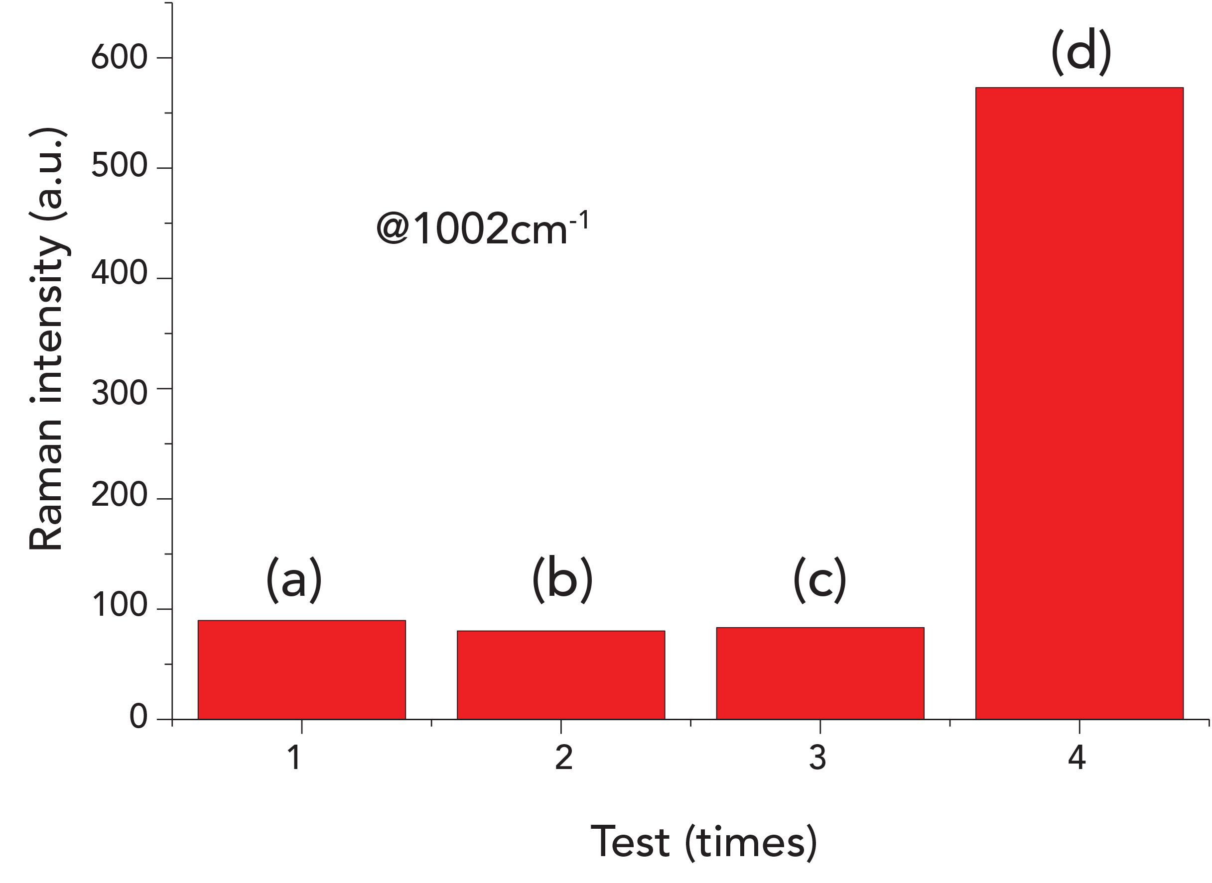
In the mixed solution, according to the actual ratio, we selected 1002 cm-1 so we can clearly see the change of the spectrum of scopolamine in the process, which is shown in Figure 6. Figure 7 displays the results in a column chart. Although the first three times have weak intensity, they are below the detection limit so they can be regarded as the absence of scopolamine. The fourth time is the success of the actual detection of scopolamine.
FIGURE 7: SERS intensity of mixture at 1002 cm-1.
Conclusions
We used simple experimental conditions to identify the spectrum of a solution mixture and utilized the disadvantageous SERS detection conditions to create a means of separating mixtures. This solves the problem that the strong adsorption component in the mixture can inhibit the weak adsorption component in the Raman spectrum. It should be noted that in the process of continuous centrifugation, the mixed solution is diluted to a certain extent. Although the relative concentration ratio of scopolamine is increasing, the concentration of scopolamine itself is decreasing, and therefore, the number of centrifugations needs to be properly controlled. The amount of silver sol used for centrifugation also requires optimization. Compared with other methods of separating mixtures, the procedure described herein is simple, and it is not only a feasible new method for the SERS detection of solution mixtures, but also for a rapid screening of new alternative drugs.
Acknowledgments
This work was supported by the National Natural Science Foundation of China( Grant No. 31871873), Inner Mongolia University for the Nationalities (Grant No. NMDYB18006), and the Inner Mongolia Autonomous Region Natural Science Foundation of China (Grant No. 2018LH08055).
Conflicts of Interest
There are no conflicts to declare.
References
(1) M.K. Bohm and H.B. Clayton, J. Adolesc. Health 66, S130 (2020).
(2) J. Valentin, T. Mateo, M. Mariana, S. Virginie, S. Anne-Marie, L. Denis, S. Anais, L. Virginie, B. Fabrice, A. Serge, and R. Gerald, Drug Test. Anal. DOI:10.1002/dta.2745 (2020).
(3) Q-T. Akram and S. Mostafa, Gene 724, 144–153 (2020).
(4) S. Kelly, Crit. Public Heath 30, 68–78 (2020).
(5) K. Julia, W. Sophia, R. Francois, S. Norbert, B. Matthias, and W. Elisa, Drug Alcohol Depend. 205, 107593 (2019).
(6) B. Valentina, C. Ciro, M. Angelo, G.I.L. Francesco, P. Matteo, and M. Icro, Heroin Addict. Relat. Clin. Probl. 21, 5–16 (2019).
(7) G. Yong-Long, Z. Yang, C. Jiang-Peng, W. Sheng-Bing, C. Xing-Hui, Z. Yan-Chun, et al., Acupunct. Med. 35(5), 366–373 (2018).
(8) L. Yixin, X. Baijuan, L. Rongrong, Y. Dan, W. Yanlin and L. Wenmei, Neuroreport 28, 654–66 (2017).
(9) Z.Denghui, Z. Dengke, D. Huiqiong, Z. Xuehui, Q Jinping, S. Hongxian, X. Xiaojun, W. Xuyi and H. W, Chin. J. Drug Depend. 15, 35–39 (2006).
(10) Z. Xuhui, W. Xuyi, L. Jun and H. Wei, Chin. J. Clin. Psychol. 19, 285–288 (2011).
(11) S. Wenwen, W. Qing, Z. Jianbin, P. Wenkai, Z. Jiawen, Y. Weiting, H. Qianyu, C. Deniz, and Z. Wenhua, Front. Psychiatry 10, 888 (2019).
(12) C. Daniel, O. Jeff, and M. Sarah, Int. J. Drug Policy 46, 146–155 (2017).
(13) C.P. Fang, S.C. Wang, H.H. Tsou, R.H. Chung, Y.T. Hsu, S.C. Liu, H.W. Kuo, T.H. Liu, A.C.H. Chen, and Y.L. Liu, J. Hum. Genet. 65, 381–386 (2020).
14) W.W. Shen, Q. Wang, J.B. Zhang, W.K. Ping, J.W. Zhang, W.T. Ye, Q.Y. Hu, D. Cerci, and W.H. Zhou, Front. Psychiatry 10, 888 (2019).
(15) L. Bao, S. Han, X.Y. Sha, H. Zhao, Y.P. Liu, D.Y. Lin, and W. Hasi, J. Spectrosc. 2019, 7458371 (2019).
(16) J. Langer, D.J. de Aberasturi, J. Aizpurua, R. A. Alvarez-Puebla, B. Auguie, J.J. Baumberg, G.C. Bazan, S.E.J. Bell, A. Boisen, et al., ACS Nano 14, 28–117 (2020).
(17) R. Pilot, R. Signorini, C. Durante, L. Orian, M. Bhamidipati, and L. Fabris, Biosensors-Basel 9, 57 (2019).
(18) B.N.J. Persson, Chem. Phys. Lett. 82, 561–565 (1981).
(19) J. Faulds, R.E. Littleford, D. Graham, G. Dent, and W.E. Smith, Anal. Chem. 76, 592–598 (2004).
(20) C. Caro, P. Quaresma, E. Pereira, et al., Nanomaterials 9, 256 (2019).
(21) J. Langer, D.J. de Aberasturi, J. Aizpurua, et al., ACS Nano 14, 28–117 (2020).
(22) N. Leopold and B. Lendl, J. Phys. Chem. B. 107, 5723–5727 (2003).
(23) C. Carlos, J.S. Maria, F. Victorino, C. Alejandro, Z. Paula and G. Francisco, Sens. Actuator B-Chem. 228, 124–133 (2016).
(24) K. Kneipp, Y. Wang, H. Kneipp, et al., Phys. Rev. Lett. 78, 1667–1670 (1997).
(25) A.C. Sara, H. Szushen, R.G. Benito, et al., Angew. Chem.-Int. Edit. 48, 5326–5329 (2009).
(26) R.A. Sulk, R.C. Corcoran and K.T. Carron, Abstr. Pap. Am. Chem. Soc. 211, 333– ANYL (1996).
(27) K.J. Si, P.Z. Guo, Q.Q. Shi. and W.L. Cheng, Anal. Chem. 87, 5263–5269 (2015).
(28) X. Lin, W.L.J. Hasi, X.T. Lou, S. Han, D.Y. Lin and Z.W. Lu, Anal. Methods 7, 3869–3875 (2015).
(29) S.Z. Weng, M.Q. Qiu, R.L. Dong, F. Wang, L.S. Huang, D.Y. Zhang and J.L. Zhao, Spectroc. Acta Pt. A-Molec, Biomolec. Spectr. 200, 20–25 (2018).
(30) H. Zhao, W. Hasi, L. Bao, Y.P. Liu, S. Han and D.Y. Lin, J. Raman Spectrosc. 49, 1469–1477 (2018).
(31) L. Gaft and L. Nagi, Opt. Mater. 30, 1739–1746 (2008).
(32) Y.Q. Qin, X.H. Ji, J. Jing, H. Liu and H.L. Wu, Colloids Surf. A 372, 172−176 (2010).
Wuliji Hasi is with the National Laboratory of Science and Technology at the Harbin Institute of Technology in Harbin, China. Lin Bao is with the National Laboratory of Science and Technology at the Harbin Institute of Technology in Harbin, China, and the College of Physics and Electronic Information at the Inner Mongolia Universities for Nationalities. Siqingaowa Han is with the Affiliated Hospital at Inner Mongolia University for the Nationalities. Direct correspondence to Wuliji Hasi at hasiwuliji19@163.com ●

AI-Powered SERS Spectroscopy Breakthrough Boosts Safety of Medicinal Food Products
April 16th 2025A new deep learning-enhanced spectroscopic platform—SERSome—developed by researchers in China and Finland, identifies medicinal and edible homologs (MEHs) with 98% accuracy. This innovation could revolutionize safety and quality control in the growing MEH market.
New Raman Spectroscopy Method Enhances Real-Time Monitoring Across Fermentation Processes
April 15th 2025Researchers at Delft University of Technology have developed a novel method using single compound spectra to enhance the transferability and accuracy of Raman spectroscopy models for real-time fermentation monitoring.
Nanometer-Scale Studies Using Tip Enhanced Raman Spectroscopy
February 8th 2013Volker Deckert, the winner of the 2013 Charles Mann Award, is advancing the use of tip enhanced Raman spectroscopy (TERS) to push the lateral resolution of vibrational spectroscopy well below the Abbe limit, to achieve single-molecule sensitivity. Because the tip can be moved with sub-nanometer precision, structural information with unmatched spatial resolution can be achieved without the need of specific labels.