A Confocal Raman Imaging Study on Emulsions
In the life sciences and bio-medical research, in the food as well pharmaceutical industry, the development of characteristic emulsions and suspensions with distinctive features play an important role.
Harald Fischer, Wolfram Ibach, Henning Dampel, and Detlef Sanchen, WITec GmbH
In the life sciences and bio-medical research, in the food as well pharmaceutical industry, the development of characteristic emulsions and suspensions with distinctive features play an important role. As the microstructure of such mixtures strongly determines the properties of final products, powerful analytical tools are required for studying the distribution of the various compounds. Confocal Raman Imaging allows detailed structural and compositional investigation. By integrating a Raman spectrometer within a state-of-the-art confocal microscope setup, Raman imaging with a spatial resolution down to 200 nm laterally and 500 nm vertically can be achieved using visible light excitation. With this setup, a complete Raman spectrum is acquired at each image pixel, typically taking between 0.7 and 100 ms. From this multi-spectrum file, an image is generated by integrating over a certain Raman line in all spectra or by evaluating the various peak properties such as peak-width, min/max analysis, or peak position. Due to the confocal arrangement, even depth profiling and 3-D imaging are possible if the sample is transparent.
Emulsion for Food Production
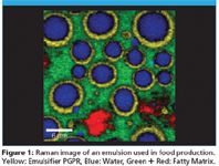
To exemplify the capabilities of this technique an emulsion used in food production was investigated using the WITec Confocal Raman Microscope alpha300 R. The emulsion which contains a fatty matrix, an aqueous phase and the emulsifier polyglycerol polyricinoleate (PGPR) was imaged with a scan range of 30 × 30 μm and 120 × 120 pixels. (14,400 spectra, 30 ms/spectrum). Figure 1 shows the color coded Raman image clearly revealing that the emulsifier PGPR (yellow) aggregates as an interface between the water droplets (blue) and the fatty matrix (green and red).

Figure 1
Sensitive Setup and Ultrafast 3D Image Acquisition
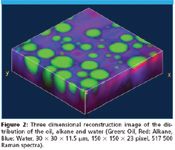
The latest spectroscopic detector technology combined with a high-throughput confocal microscope are the crucial points in the recent improvements in sensitivity allowing acquisition times for a single Raman spectrum to be reduced to 0.7 ms. Using such a sensitive setup can also be an advantage when performing measurements on delicate and precious samples requiring the lowest possible levels of excitation power. In the following study, the ultrafast Confocal Raman Imaging capabilities of the alpha300 R were used to analyze an oil-alkane-water emulsion three dimensionally. In a volume of 30 × 30 ×11.5 μm, 23 confocal Raman scans were acquired in z-direction leading to 23 Raman images each consisting of 150 × 150 pixel (22 500 spectra). The total acquisition time for one image was 60 s resulting in 23 min for the acquisition of the complete stack (517 000 Raman spectra). Using a 3-D reconstruction software, a three dimensional image of the distribution of the three compounds can be created as shown in Figure 2 (green: oil, red: alkane, blue: water).

Figure 2
Conclusion
It was demonstrated that confocal Raman imaging provides the ability to non-invasively image the chemical properties of emulsions three dimensionally with the highest resolution.

WITec GmbH
Lise-Meitner-Str. 6, 89081 Ulm, Germany
tel. +49 (0) 731 140 700; fax +49 (0) 731 140 70 200
Website: www.witec.de

Thermo Fisher Scientists Highlight the Latest Advances in Process Monitoring with Raman Spectroscopy
April 1st 2025In this exclusive Spectroscopy interview, John Richmond and Tom Dearing of Thermo Fisher Scientific discuss the company’s Raman technology and the latest trends for process monitoring across various applications.
A Seamless Trace Elemental Analysis Prescription for Quality Pharmaceuticals
March 31st 2025Quality assurance and quality control (QA/QC) are essential in pharmaceutical manufacturing to ensure compliance with standards like United States Pharmacopoeia <232> and ICH Q3D, as well as FDA regulations. Reliable and user-friendly testing solutions help QA/QC labs deliver precise trace elemental analyses while meeting throughput demands and data security requirements.