Discrimination of Bacterial Growth Media Using Portable Raman Spectroscopy with Background Fluorescence Subtraction
The feasibility of identifying different types of bacterial growth media using a handheld Raman instrument with a proprietary baseline correction algorithm is investigated.
We investigated the feasibility of identifying different types of bacterial growth medium using a handheld Raman instrument with a proprietary baseline correction algorithm. Our data suggest that this instrument is capable of differentiating multiple types of growth medium and readily detects the change in the Raman signal when bacteria are present in the growth medium. These findings support the notion that a handheld Raman spectrometer may be used to distinguish highly-fluorescent biological samples without the use of surface-enhanced Raman scattering.
In the field of forensic sciences, handheld Raman spectrometers are used by the military and first responders for the rapid identification of unknown substances (1–3). These unknown materials include toxic industrial chemicals (TICs), toxic industrial materials (TIMs), chemical warfare agents, explosives, narcotics, pharmaceutical compounds, and plastics. However, Raman spectroscopy has historically been problematic for the identification of biological samples, such as bacteria, growth media, and tissues. Sample fluorescence typically overwhelms Raman scattering by several orders of magnitude, resulting in a high background and poor spectral matching (4).
Two general methodologies have been used to detect weak Raman signals from biological organisms and materials: surface-enhanced Raman scattering (SERS) and fluorescence background subtraction (also known as baseline correction). SERS is a technique that boosts the signal of Raman-active molecules when measured on customized metal surfaces, typically gold, silver, and copper (5,6). The increase in Raman signal on these metals is often dramatic, usually on the order of 104–106, but it can be as high as 1011 (7,8). Therefore, increasing the Raman signal over background fluorescence allows the Raman spectra to be discretely visible. In fact, a number of publications have shown that different types of bacteria and growth media have unique SERS spectra, thereby establishing SERS as a potential pathogen diagnostic tool despite the intrinsic fluorescence of the sample (9–22).
Fluorescence background subtraction is a second option for "teasing out" a weak Raman signal from fluorescent biological samples. In contrast to SERS, fluorescence background subtraction does not increase Raman signal during acquisition; it differentiates it computationally by deducting the background fluorescence from the Raman signal post-acquisition. Several computational methods have been developed for background subtraction from Raman spectra, including background estimation using computational geometry (23), principal component analysis (24), wavelet transformation (25–30), polynomial fitting (4,31–36), frequency-domain filtering (37), and first-order and second-order differentiation (38,39).
In this study, we investigated the feasibility of fluorescence background subtraction using a handheld spectrometer to differentiate several bacterial growth media, specifically in the presence and absence of bacteria. As SERS is often not practical for first responders and military personnel (because of time constraints, limited dexterity in personal protective equipment, and ease of use), we tested a portable Raman spectrometer that utilizes a proprietary algorithm to subtract background fluorescence. This baseline correction capability may allow for the interrogation of biologic materials that would otherwise "swamp" traditional handheld Raman instruments used in forensic analyses.
Experimental Conditions
Handheld Raman Spectrometers
A NanoRam handheld spectrometer was obtained from B&W Tek, Inc. The operation presets were as follows: excitation wavelength = 785 nm, laser power = 90% (270 mW), number of hits displayed = 3, and spectral range = 176–2900 cm-1. For sample analysis, the spectrometer was fitted with the vial adaptor for 4-mL glass vials.
Bacterial Growth Media
Four types of bacterial growth media were analyzed for this study: LB-Miller broth, tryptic soy broth (TSB), brain heart infusion (BHI) broth, and brain heart infusion-cysteine (BHIc) broth. All base media were purchased from Becton Dickinson and prepared according to the manufacturer's recommendations. BHIc medium required additional supplementation as reported previously (40). Briefly, 1 L of BHI medium (37 g/L) containing cysteine (1 g/L) was sterilized following the manufacturer's recommendations. The following supplements were then prepared and added before use: β-nicotinamide adenine dinucleotide (β-NAD) (1 mg/mL — dissolve 200 mg β-NAD in 200 mL of deionized water, filter sterilize using a 0.2-μm filter, store at 4 °C), heme-histidine (1 mg/mL — add 200 mg of L-histidine, 200 mg of hemin-HCl and 4 mL of 1 N NaOH to 196 mL of deionized water; microwave briefly to dissolve; filter sterilize; store at 4 °C away from light), and glucose (25 mL, 20% w/v). All BHIc supplements were purchased from Sigma-Aldrich.
Raman Spectroscopy of Bacterial Growth Media
For Raman spectroscopy of bacterial media, 1 mL of LB-Miller broth, TSB, BHI, and BHIc media were transferred to separate 4-mL screw-cap glass vials (Fisher). Each medium sample was then added to a "User Defined Library." For the handheld spectrometer, a unique Raman spectrum for each medium was captured and added to the library. After the user library was established, each medium vial was subsequently analyzed an additional 10 times (n = 10) to determine if the spectrometer could distinguish between the different growth media. The accuracy rate and hit quality index (HQI) value (that is, confidence score) for the 10 iterations were then recorded.
Raman Spectroscopy of Bacterial Cultures
Overnight cultures were set up in each medium with three different organisms: Escherichia coli C3000 (obtained from Oklahoma State University), Shigella dysenteriae (BEI number NR-520), and Vibrio cholerae (BEI number NR-144). A loopful of frozen glycerol stock was used to inoculate 5 mL of each medium in a 50-mL vented flask (Cellstar) and cultured for 18 h at 37 °C with shaking. Then, 1 mL of each sample was analyzed (n = 10) and matched against the User Defined Library. Scans were recorded as a "match" if the instrument correctly identified the medium as the top match and an HQI ≥ 90. If there were no hits, or the instrument did not correctly identify the compound, it was recorded as a "no-match." The accuracy rate and HQI value (that is, confidence score) for the 10 iterations were then recorded. The bacterial titer for each culture was calculated by performing serial 10-fold dilution and spotted onto tryptic soy agar (TSA) culture plates (20 μL/spot) in triplicate for each sample and dilution. Plates were incubated under aerobic conditions at 37 °C and the colony forming units (cfu) were calculated. The titers for Escherichia coli in LB, TSB, and BHI were 5 × 10⁸, 9.5 × 10⁸, and 4.5 × 10⁸ cfu/mL, respectively. The titers for Shigella dysenteriae in LB, TSB, and BHI were 3 × 10⁶, 1 × 10⁸, and 8 × 10⁷ cfu/mL, respectively. The titers for Vibrio cholerae in LB, TSB, and BHI were 9.0 × 10⁷, 1.3 × 10⁸, and 2.5 × 10⁸ cfu/mL, respectively.
Raman Spectroscopy of Filtered Bacterial Cultures (Spent Culture Medium)
Overnight cultures were started in LB, BHI, and TSB media with Escherichia coli C3000, Shigella dysenteriae, and Vibrio cholerae as described previously. The following day, bacteria were removed from the cultures using a 50-mL Millipore sterile disposable vacuum filtration unit with a 0.22-μm PES membrane. The resulting filtered media samples were then analyzed (n = 10) by the handheld Raman instrument in replicate experiments (three separate cultures).
Data Analysis
All data scans were downloaded from the handheld Raman instrument into NanoRam ID Client v3.10 software. In ID Client, each overlay (sample scan and library match) was reviewed and a representative scan was chosen for each data set. Screen shots of the data scans were then captured and imported into Photoshop CS5 software (Adobe) where they were cropped and organized into figures. All graphing was performed in Microsoft Excel software. A notable limitation of the handheld instrument is displaying a library spectrum with a sample spectrum that has a low HQI value (does not match). By default, the instrument displays the sample spectrum overlaid with the library spectrum that has the highest HQI value (the best match). Therefore, to have the instrument overlay a library spectrum with a dissimilar sample spectrum (for the purpose of demonstration in the figure), we created some user libraries with only a single medium entry (for example, unadulterated BHI was entered into a user library and used to match bacterial cultures propagated in BHI; even though the Raman spectra were dramatically different, the instrument would overlay the library spectrum of BHI alone with the sample spectrum of BHI and the bacteria).
Results
To investigate the utility of using a handheld Raman spectrometer for identifying bacterial medium (Figure 1), we tested four different types of growth medium with increasing complexity: LB-Miller Broth (LB), Tryptic Soy Broth (TSB), Brain-Heart Infusion Broth (BHI), and a complex derivative of BHI used for culturing Francisella tularensis (BHIc). The ingredients for each medium are listed in Table I.
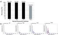
Figure 1: (a) Measuring the accuracy of the portable spectrometer on four types of bacterial growth medium: LB, TSB, BHI, and BHIc. Scans were recorded as a "match" if the instrument correctly identified the medium as the top match with an HQI value ⥠90. Scans that exceeded this average value are shown in black, while scans below this threshold value are show in grey. (b) Representative overlays of the baseline-corrected sample spectrum (blue line) and the library spectrum (red line) are shown for each of the four types of growth medium.
The accuracy rate (%) was determined by interrogating 10 sample replicates for each type of medium. Scans were recorded as a "match" if the instrument correctly identified the medium as the top match in the library, and the HQI value was ≥ 90. The HQI value indicates how well the unknown scan matches to the library scan; an HQI of 100 indicates an identical match while lower values indicate the dissimilarity between scans (41). As shown in Figure 1a, the NanoRam spectrometer was able to match LB, TSB, and BHI media with 100% accuracy with HQI values ≥ 94. BHIc, the most complex medium in the study, matched with 80% accuracy with an average HQI value of 78. Figure 1b shows a representative spectrum from each medium analyzed. Scans for LB, TSB, and BHI were nearly identical to the library scans (blue tightly fit to red). However, the spectrum for the BHIc medium was more variable compared to the library spectrum (red visible with no blue), indicating that the match was of lower quality. These findings suggest that the NanoRam is likely able to match to a variety of bacterial growth media using its proprietary background subtraction algorithm. However, analyzing highly-complex medium such as BHIc results in lower HQI values.
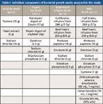
Table I: Individual components of bacterial growth media analyzed in this study
After determining that the instrument was able to distinguish between different types of growth medium, we investigated whether culturing three types of enteric bacteria (Escherichia coli, Shigella dysenteriae, and Vibrio cholerae) dramatically affected the Raman spectra compared to culture medium alone. We also assessed whether these three types of bacteria could be visually distinguished from one another by their subtracted Raman spectra (as a proof-of-principle). Escherichia coli, Shigella dysenteriae, and Vibrio cholerae were cultured in LB, TSB, and BHI; these cultures were then analyzed by the NanoRam spectrometer to compare spectra of broth alone and broth containing bacteria. Interestingly, both the Escherichia coli and Shigella dysenteriae cultures in LB matched, with 100% accuracy, to the library scans of LB medium alone (Figure 2a). A similar finding was observed by Premasiri and colleagues (18), who noted that "media containing extracts from other micro-organisms such as yeast extract" showed a similar appearance to SERS spectra of bacterial cells, an observation consistent with culturing these bacteria in LB (a yeast extract base). By contrast, spectra from cultures of Escherichia coli, Shigella dysenteriae, and Vibrio cholerae in TSB and BHI were notably different than those of medium alone, and did not match the library spectra (Figure 2a). Figure 2b shows the Raman spectra of LB, TSB, and BHI alone (reference scans, first row) compared to the three enteric bacteria cultured in these media (rows 2–4). Note that the red library scan, for each of the representative Raman plots, was from pure medium alone (LB, TSB, or BHI); the blue sample scan represents the change in the Raman spectrum from the bacterial culture relative to medium alone. Although Escherichia coli and Shigella dysenteriae cultured in LB had similar scans relative to LB alone, all three bacteria showed unique background-subtracted Raman spectra in TSB and BHI, suggesting that it may be feasible to distinguish between multiple types of bacteria cultured in a variety of growth media using a handheld Raman spectrometer without using SERS.
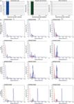
Figure 2: (a) Culturing Escherichia coli, Shigella dysenteriae, and Vibrio cholerae in LB, TSB, or BHI markedly decreased the accuracy rate of the portable spectrometer to match to the growth medium spectra in the library. (b) Representative overlays of the baseline-corrected sample spectrum (blue line) and the library spectrum (red line) are shown for each of the three types of growth medium in the presence of Escherichia coli, Shigella dysenteriae, and Vibrio cholerae. The medium alone scan (reference) is unadulterated growth medium matched to itself.
Next, we investigated whether the robust Raman peaks observed in the bacterial cultures from Figure 2 were caused exclusively by the presence of the bacteria, or whether the Raman-active components in the growth medium had also changed during bacterial growth. We hypothesized that if the dramatic changes in Raman spectra were due to bacteria alone, filtering out these bacteria from the spent culture medium would restore the ability of the handheld Raman spectrometer to match to the "medium alone" library scans, specifically for TSB and BHI (bacterial cultures that had a 0% match rate for all three bacteria). To test this hypothesis, overnight bacterial cultures were grown to saturation. Cultures were then filtered through a 0.22-μm membrane to remove bacteria; spent culture media were then analyzed using the handheld Raman spectrometer (Figure 3a). Filtering out the bacteria largely restored the ability of the instrument to match TSB spent culture medium to the original TSB library scan (with an HQI value ≥ 90), whereas the BHI spent culture medium did not match to the library scan at the original HQI threshold. However, most of the BHI scans had HQI values ranging from 75 to 89, suggesting that the BHI spent culture medium was similar to unadulterated BHI. Representative scans also supported the notion that the spent culture medium had minor differences in their Raman spectra compared to the original growth medium, although these differences were enough to drop the HQI value below our threshold for a match (Figure 3b). These data suggest that the bacteria in the culture medium are primarily responsible for the dramatic change in the background-subtracted Raman spectra compared to medium alone.
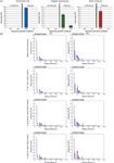
Figure 3: (a) Removing bacteria from culture medium partially restores the ability of the handheld spectrometer to match to the growth medium spectra in the library. (b) Representative overlays of the baseline-corrected spent culture medium (blue line) and the library spectrum (red line) are shown for TSB and BHI after Escherichia coli, Shigella dysenteriae, and Vibrio cholerae have been filtered from the culture.
Discussion and Conclusion
We investigated whether a handheld Raman instrument that subtracts background fluorescence was able to identify increasingly complex bacterial growth medium, with and without bacteria. We found that the instrument was effective in identifying several types of traditional growth medium at an HQI value of ≥ 90, but was less accurate with complex specialty mediums, such as BHIc. In addition, culturing bacteria in these media resulted in markedly different Raman spectra compared to media alone, suggesting that it may be feasible to distinguish between multiple types of bacteria cultured in a variety of growth media if an on-board reference library is generated first. We also noted variability in the intensity and position of peaks in replicate spectra for each condition, suggesting that if a reference library were to be generated, each library spectrum would need to be an average of multiple scans (ideally).
Two recent studies have discussed the use of SERS to differentiate bacterial growth medium and different species of bacteria (that is, by boosting the Raman signal during acquisition) (16,18). We show in this study that growth medium and bacteria could also be differentiated using a fluorescence background subtraction algorithm post-acquisition. This is particularly relevant for first responders and military personnel who use handheld Raman spectrometers to identify chemicals (TICs), toxic industrial materials (TIMs), chemical warfare agents, explosives, and narcotics, but historically struggle with "bio" materials because of their high background fluorescence. Therefore, future studies will build upon these initial findings and investigate the ability of handheld Raman spectrometers to correctly identify a multitude of crude bacterial cultures, purified bacterial cells (washed), as well as dried spores.
References
(1) H. Markert, J. Rings, N. Campbell, and K. Grates, N. F. S. T. Center, Ed. (National Institute of Justice, Largo, Florida, 2011), pp. 1–8.
(2) W. Smith, P. White, C. Rodger, and G. Dent, in Handbook of Raman Spectroscopy: From the Research Laboratory to the Process Line, I.R. Lewis and H. Edwards, Eds. (Marcel Dekker, New York, New York, 2001).
(3) V. Otieno-Alego and N. Speers, in Infrared and Raman Spectroscopy in Forensic Science, J.M. Chalmers, H. Edwards, and M. Hargreaves, Eds. (John Wiley & Sons Ltd, West Sussex, UK, 2012).
(4) C.A. Lieber and A. Mahadevan-Jansen, Appl. Spectrosc. 57, 1363 (2003).
(5) M. Culha, B. Cullum, N. Lavrik, and C. Klutse, J. Nanotechnol. 2012, 15 (2012).
(6) B. Sharma, R. Frontiera, A. Henry, E. Ringe, and R. Van Duyne, Mater. Today 15, 16 (2012).
(7) K. Kneipp, H. Kneipp, I. Itzkan, R. R. Dasari, and M.S. Feld, Chem. Rev. 99, 2957 (1999).
(8) K. Kneipp, H. Kneipp, I. Itzkan, R.R. Dasari, and M.S. Feld, Curr. Sci. 77, (1999).
(9) H. Chu, Y. Huang, and Y. Zhao, Appl. Spectrosc. 62, 922 (2008).
(10) J.A. Guicheteau, K. Gonser, and S.D. Christesen, A.P.G. National Research Council Associate at the U.S. Army ECBC Research Lab, Ed. (U.S. Government, Edgewood, 2004).
(11) R.M. Jarvis, A. Brooker, and R. Goodacre, Anal. Chem. 76, 5198 (2004).
(12) R.M. Jarvis and R. Goodacre, Anal Chem. 76, 40 (2004).
(13) M. Kahraman, M.M. Yazici, F. Sahin, and M. Culha, J. Biomed. Opt. 12, 054015 (2007).
(14) M. Kahraman, M.M. Yazici, F. Sahin, and M. Culha, Langmuir 24, 894 (2008).
(15) T.T. Liu et al., PLoS One 4, e5470 (2009).
(16) N.E. Marotta and L.A. Bottomley, Appl. Spectrosc. 64, 601 (2010).
(17) I.S. Patel, W.R. Premasiri, D.T. Moir, and L.D. Ziegler, J. Raman Spectrosc. 39, 1660 (2008).
(18) W.R. Premasiri, Y. Gebregziabher, and L.D. Ziegler, Appl. Spectrosc. 65, 493 (2011).
(19) W.R. Premasiri et al., J. Phys. Chem. B 109, 312 (2005).
(20) W.R. Premasiri, D.T. Moir, M.S. Klempner, and L.D. Ziegler, Eds., New Approaches in Biomedical Spectroscopy, (Oxford University Press, New York, 2007).
(21) A. Sengupta, M.L. Laucks, and E.J. Davis, Appl. Spectrosc. 59, 1016 (2005).
(22) L. Zeiri, B.V. Bronk, Y. Shabtai, J. Eichler, and S. Efrima, Appl. Spectrosc. 58, 33 (2004).
(23) N. Kourkoumelis, A. Polymeros, and M. Tzaphlidou, Spectrosc. Int. J. 27, 311 (2012).
(24) T. Hasegawa, J. Nishijo, and J. Umemura, Chem. Phys. Lett. 317, 642 (2000).
(25) T.T. Cai, D. Zhang, and D. Ben-Amotz, Appl. Spectrosc. 55, 1124 (2001).
(26) C. Camerlingo, F. Zenone, G.M. Gaeta, R. Riccio, and M. Lepore, Meas. Sci. Technol. 17, 298 (2006).
(27) Y. Hu et al., Chemom. Intell. Lab. Syst. 85, 94 (2007).
(28) J. Li, L.P. Choo-Smith, Z. Tang, and M.G. Sowa, J. Raman Spectrosc. 42, 580 (2011).
(29) P.M. Ramos and I. Ruisánchez, J. Raman Spectrosc. 36, 848 (2005).
(30) Z. Zhang et al., J. Raman Spectrosc. 41, 659 (2010).
(31) B.D. Beier and A.J. Berger, Analyst 134, 1198 (2009).
(32) A. Jirasek, G. Schulze, M.M. Yu, M.W. Blades, and R.F. Turner, Appl. Spectrosc. 58, 1488 (2004).
(33) M.N. Leger, A.G. Ryder, Appl. Spectrosc. 60, 182 (2006).
(34) V. Mazet, C. Carteret, D. Brie, J. Idier, and B. Humbert, Chemom. Intell. Lab. Syst. 76, 121 (2005).
(35) T.J. Vickers, R.E. Wambles, and C.K. Mann, Appl. Spectrosc. 55, 389 (2001).
(36) J. Zhao, H. Lui, D.I. McLean, and H. Zeng, Appl. Spectrosc. 61, 1225 (2007).
(37) P.A. Mosier-Boss, S.H. Lieberman, and R. Newbery, Appl. Spectrosc. 49, 630 (1995).
(38) A. O'Grady, A.C. Dennis, D. Denvir, J.J. McGarvey, and S.E. Bell, Anal. Chem. 73, 2058 (2001).
(39) D. Zhang and D. Ben-Amotz, Appl. Spectrosc. 54, 1379 (2000).
(40) P. Mc Gann et al., J. Microbiol Methods 80, 164 (2010).
(41) J. Kauffman, J.D. Rodriguez, and L.F. Buhse, Am. Pharm. Rev. 14, (2011).
Jessica A. Randall and Mathew G. Lyman are with the Chemical, Biological, Radiological, Nuclear, and Explosives Division at Oklahoma State University – University Multispectral Laboratory in Ponca City, Oklahoma. Direct correspondence to: mlyman@okstate-uml.org
AI-Powered SERS Spectroscopy Breakthrough Boosts Safety of Medicinal Food Products
April 16th 2025A new deep learning-enhanced spectroscopic platform—SERSome—developed by researchers in China and Finland, identifies medicinal and edible homologs (MEHs) with 98% accuracy. This innovation could revolutionize safety and quality control in the growing MEH market.
New Raman Spectroscopy Method Enhances Real-Time Monitoring Across Fermentation Processes
April 15th 2025Researchers at Delft University of Technology have developed a novel method using single compound spectra to enhance the transferability and accuracy of Raman spectroscopy models for real-time fermentation monitoring.
Nanometer-Scale Studies Using Tip Enhanced Raman Spectroscopy
February 8th 2013Volker Deckert, the winner of the 2013 Charles Mann Award, is advancing the use of tip enhanced Raman spectroscopy (TERS) to push the lateral resolution of vibrational spectroscopy well below the Abbe limit, to achieve single-molecule sensitivity. Because the tip can be moved with sub-nanometer precision, structural information with unmatched spatial resolution can be achieved without the need of specific labels.