Tissue Raman Measurement at 1064 nm
Application Notebook
Biological tissues and other materials often autofluoresce at near-infrared wavelengths, prohibiting Raman acquisition. New, dispersive Raman systems at 1064 nm allow fluorescence-free measurement in similar integration times.
Biological tissues and other materials often autofluoresce at near-infrared wavelengths, prohibiting Raman acquisition. New, dispersive Raman systems at 1064 nm allow fluorescence-free measurement in similar integration times.
Fluorescence is much more likely and intense at short wavelengths where energies are more apt to cause an electronic transition. Yet even at near-IR wavelengths like 785 or 830 nm, many substances still fluoresce, sometimes prohibiting Raman spectral acquisition. For those users who require longer wavelengths such as 1064 nm, the only available option has been FT-Raman, which is typically noisier and slower than dispersive Raman systems. But now, BaySpec's new dispersive 1064 nm Raman spectrometer family of instruments offers a turn-key solution that combines the speed, sensitivity, and rugged design of traditional dispersive Raman instruments with the fluorescence avoidance of traditional FT-Raman instruments.
Experimental Conditions
A variety of animal tissues obtained from a local market were interrogated using two BaySpec benchtop systems: the RamSpec™ 785 and the RamSpec™ 1064. Both systems utilized filtered fiber probes designed for each respective wavelength. Acquisition times were 30 s for both systems. Power was set at 50 mW for the 785 nm measurements, and 150 mW for the 1064 nm measurements.
Results
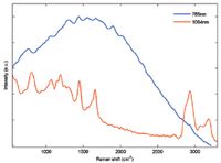
Pigmented and porphyrin-rich tissues such as kidney are often too fluorescent to be measured, even at 785 nm; see Figure 1. But using 1064 nm, a clear Raman spectrum relatively devoid of fluorescence background is generated using the same integration time. Additionally, because of the extended quantum efficiency of the InGaAs detector, high wavenumber features (C–H, O–H, and N–H stretching modes) are also simultaneously captured with the same laser.

Figure 1: Kidney (porcine) is highly fluorescent at 785 nm, preventing extraction of a usable Raman signal. At 1064 nm, however, this fluorescence interference is largely avoided and clear Raman bands are evident.
In addition to allowing users the ability to measure Raman spectra from highly fluorescent samples, the dispersive 1064 nm Raman systems also reduce the stringent sampling conditions necessary at shorter wavelengths. For example, 785-nm Raman measurement through vials, cuvettes, or under cover slips often requires that these materials be made of fused silica, quartz, or calcium fluoride for their reduced fluorescence, all of which cost considerably more than bulk glass materials. However, even inexpensive glass sample vials can be used for 1064-nm measurement of weak Raman scatterers such as tissue, owing to the fluorescence avoidance of the longer wavelength approach.
One of the lingering concerns about the use of 1064-nm Raman is the reduced Raman scattering cross-section. As compared to 785-nm Raman, this cross-section, indeed, reduces approximately 3.4×. However, according to permissible standards for human tissue (skin) exposure, the maximum power level that can be tolerated at 1064 nm is approximately 3.4× higher than the power permissible at 785 nm (1). So, even in photosensitive samples such as biological tissue, the physical reduction in Raman efficiency can be totally compensated by increased laser power.
Conclusions
Based on these experiments, 1064-nm dispersive Raman will provide a viable new option for those users who are studying highly fluorescent tissues, desire to measure multiple samples in simple glass containers, or those users who are interested in simultaneous fingerprint and high wavenumber spectral acquisition. Future studies will certainly evidence further advantages of this approach, as compared to shorter wavelength (785 or 830 nm) Raman approaches or FT-Raman systems.
References
(1) National Institute of Standards and Technology, Z136.1, 76–77, (2007).
BaySpec, Inc.
1101 McKay Drive, San Jose, CA 95131
tel. (408) 512-5928, fax: (408) 512-5929
Website: www.bayspec.com

Thermo Fisher Scientists Highlight the Latest Advances in Process Monitoring with Raman Spectroscopy
April 1st 2025In this exclusive Spectroscopy interview, John Richmond and Tom Dearing of Thermo Fisher Scientific discuss the company’s Raman technology and the latest trends for process monitoring across various applications.
A Seamless Trace Elemental Analysis Prescription for Quality Pharmaceuticals
March 31st 2025Quality assurance and quality control (QA/QC) are essential in pharmaceutical manufacturing to ensure compliance with standards like United States Pharmacopoeia <232> and ICH Q3D, as well as FDA regulations. Reliable and user-friendly testing solutions help QA/QC labs deliver precise trace elemental analyses while meeting throughput demands and data security requirements.