Biological Mapping with LIBS
Vincent Motto-Ros of Lyon 1 University, in Lyon, France, is combining the ability of atomic spectroscopy techniques to detect and quantify metals with the mapping approaches most often used with molecular techniques. He has combined laser-induced breakdown spectroscopy (LIBS) with electron microscopy to map the metals and metallic nanoparticles in biological tissue, as a way of studying the update and clearance of these materials by biological systems. In this interview, he discusses his work applying LIBS to biological analysis, including the methods, advantages, and future directions.
Vincent Motto-Ros of Lyon 1 University, in Lyon, France, is combining the ability of atomic spectroscopy techniques to detect and quantify metals with the mapping approaches most often used with molecular techniques. He has combined laser-induced breakdown spectroscopy (LIBS) with electron microscopy to map the metals and metallic nanoparticles in biological tissue, as a way of studying the update and clearance of these materials by biological systems. In this interview, he discusses his work applying LIBS to biological analysis, including the methods, advantages, and future directions.
In recent years, many researchers have been advancing the application of molecular spectroscopy techniques, such as Raman and infrared (IR) spectroscopy, to image biological tissues, such as for the analysis of tumors. Atomic spectroscopy techniques such as inductively coupled plasma-mass spectrometry (ICP-MS), on the other hand, are being used extensively to study metals and metallic nanoparticles in biological matrices. Vincent Motto-Ros of the Light and Matter Institute at Lyon 1 University, in Lyon, France, is combining the ability of atomic techniques to detect and quantify metals with the scanning approaches most often used with molecular techniques. He has used laser-induced breakdown spectroscopy (LIBS) to image metals and metallic nanoparticles in biological tissue, as a way of studying the uptake and clearance of these materials by biological systems. He recently spoke to us about this work.
You are developing LIBS techniques for the imaging analysis of metallic nanoparticles and other metals in biological tissue. How does LIBS imaging work?
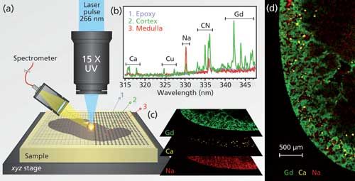
In LIBS imaging, laser-induced plasma are generated continuously while scanning the sample surface over the region of interest. Elemental images are obtained, in a pixel-by-pixel manner, after extracting the intensity of the interesting species (atoms, ions, or molecules) from each recorded spectrum (Figure 1).

Figure 1: (a) Principle of LIBS imaging instrument with the major components, including the microscope objective used to focus the UV laser pulse, the motorized platform supporting the sample, and the optical detection system fiber-connected to the spectrometer. (b) Example of single-shot emission spectra in the 315–345 nm spectral range recorded in three different regions of the mouse kidney sample indicated in (a) with emission lines of calcium (Ca), sodium (Na), and gadolinium (Gd). (c) Relative-abundance images of Gd (green), Ca (yellow), and Na (red) represented in a false color scale. (d) Multielemental biodistributions in a coronal murine kidney section, 24 h after Gd-based nanoparticle administration (spatial resolution of 10â µm).
In a recent study in mice, you used LIBS to investigate the renal clearance of gadolinium-based nanoparticles that are being studied for their potential as both diagnostic tools and therapeutics (1). What exactly were you looking for?
Nanomaterials represent a huge potential for future medical applications, but the characterization of these attractive candidates within biological tissue remains very challenging, especially because of their small size. Any modification applied to such small nanoparticles, such as fluorescence labeling, may indeed modify their shape, size, or charge, and therefore affect their biodistribution. In this work, we wanted to study the renal biodistribution of gadolinium-based nanoparticles with the aim to show the potential of LIBS for label-free imaging of nanoparticles, and metals more generally, in their biological environment.
What benefits can LIBS offer for such studies compared to existing elemental methods?
The LIBS approach is the only optical technique able to image elements with parts-per-million sensitivity and a resolution in the range of a few micrometers. The simplicity of the setup endows LIBS with a series of advantages including an all-optical design compatible with standard optical microscopy, reduced cost, and operation in ambient atmosphere. In addition, although LIBS is not as sensitive as laser-ablation ICP-MS (LA-ICP-MS), which has a sensitivity of
< 500 ppb, or as high-resolving as micro X-ray fluorescence spectrometry (< 1 µm), it has a fast operating speed, which can be up to 100 times faster than other techniques. These advantages enable LIBS imaging to provide large-scale images over entire organs (> 1 cm²) with a cellular scale resolution (~10 µm) and in reasonable time periods.
How did you optimize the balance between spatial resolution and detection sensitivity?
Achieving an adequate balance between spatial resolution and detection sensitivity is indeed one of the critical points for setting up a LIBS imaging instrument. In LIBS, sensitivity and the resolution are tightly linked. Resolution is governed by the size of the ablation craters, whereas sensitivity relies on the amount of vaporized material and on the excitation capability of the laser pulse. For example, improving the resolution (using for example an objective with a higher magnification) may dramatically lower the sensitivity. Considering our given experimental configuration (UV laser pulse, x15 magnification objective, collection system, and so on) the best performance, both in terms of signal-to-noise ratio and crater size, was obtained for a pulse energy of 800 µJ.
In this study, the kidney samples were embedded in epoxy before analysis. Would it be possible to do this kind of analysis on fresh tissue?
Yes, our first studies were carried out on fresh tissue slices (2,3). However, mastering the laser ablation of such soft materials is difficult and the accessible resolution is quite poor (~100 µm). The use of epoxy-embedded samples offers better ablation control compared with fresh tissue, which greatly improves the sample’s mechanical stability, the ablation shot-to-shot repeatability, and thus the accessible resolution.
How were the elements of interest quantified using the imaging technique?
A standard quantification methodology was used. Standards were made in epoxy resin with graduated concentration of the elements of interest (Gd, Si, Na, Fe). Calibration curves were then built for each element and the relative-abundance images were subsequently transformed into quantitative-abundance images.
What limits of detection were you able to attain for the various elements you measured?
We typically obtained detection limits of 10 ppm for Gd, 2 ppm for Si, and 5 ppm for Na, values estimated from single-shot measurements.
What were the initial results in this study, particularly as you studied the renal clearance of the nanoparticles over time?
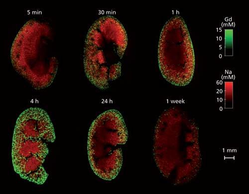
These LIBS experiments made it possible to image the components of the nanoparticles (Gd and Si) throughout the entire kidney and provided evidence of both their rapid renal uptake and their elimination. We also imaged elements naturally present in the tissue such as Na, Ca, Fe, Cu, P, and Mg. The kinetics of nanoparticle elimination was evaluated from kidney samples collected at various times after nanoparticle administration (Figure 2). These results highlight the appropriate elimination of nanoparticles from the organism, which is a fundamental necessity for the clinical development of such agents.

Figure 2: Quantitative imaging of Gd and Na in kidney coronal sections showing the fast renal uptake and elimination of nanoparticles. Mouse kidney samples were collected at various times after nanoparticle administration (ranging from 5 min to 1 week).
Overall, what did your study show about the suitability of LIBS to this type of work?
The LIBS imaging capability is very complementary to conventional methods for biological research, such as transmission electron microscopy or immunohistochemistry. Importantly, this work has drawn the attention of biologists working in the field of nanotechnology. They are seeing in LIBS a promising approach for preclinical studies with the idea to provide the biodistribution of nanomaterials at the scale of an entire organ. Since doing this study, we have worked on several applications where LIBS imaging has been used among other techniques to evaluate the biological activity and toxicity of various types of nanoparticles (4–6).
How large an area did you sample, and how long did it take? How feasible is this method for mapping larger samples?
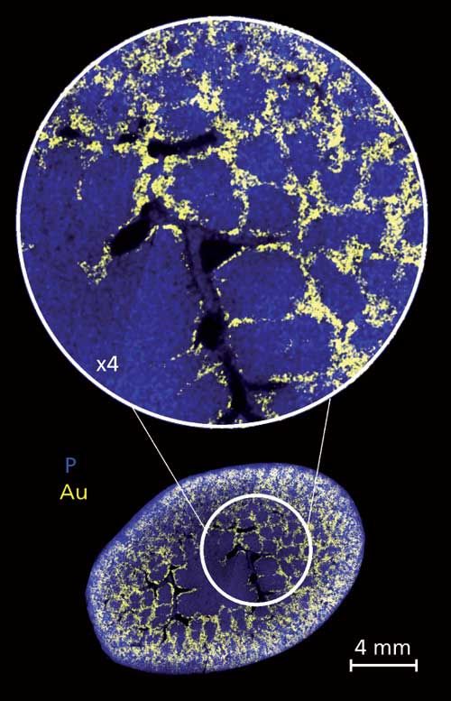
Imaging large areas by LIBS is not technically difficult as long as the sample is perfectly flat (or alternatively the instrument has to be equipped with an autofocus system). The scanning speed is critical, however, because recording high-definition images with a large number of pixels can take a considerable amount of time. All the images shown above were obtained using an instrumentation operating at 10 Hz. With this acquisition rate, 36,000 pixels were recorded within 1 h. This year, we updated our instruments to increase the scanning rate to 100 Hz. This obviously provides more comfortable conditions for analyzing larger samples. An example is shown in Figure 3. This image of 500,000 pixels represents a rat kidney section (~2 cm²) and was recorded in about 80 min at 100 Hz.

Figure 3: High-definition image showing the biodistribution of gold-based nanoparticles in a rat kidney. This image of around 500,000 pixels was recorded with a resolution of 20 µm and covers a surface of about 2 cm².
What are your next steps in this work?
As already mentioned, we are working on the improvement of the acquisition rate. We should be able to make our first kilohertz tests in a couple of weeks. In addition, we are also investigating the three-dimensional capability of the technique with the aim both to provide a global organ distribution of metals and to focus on specific regions of the tissue with cellular-scale resolution. At the present time, we are also working on samples with more complex architectures such as the brain, lungs, or tumors, and with other types of metal-based nanoparticles (gold, silver, platinum, and so on).
References
- L. Sancey, V. Motto-Ros, B. Busser, S. Kotb, J.M. Benoit, A. Piednoir, F. Lux, O. Tillement, G. Panczer, and J. Yu, Scientific Reports4, 6065 (2014).
- V. Motto-Ros et al., Spectrochim. Acta, Part B83, 168–174 (2013).
- V. Motto-Ros et al., Appl. Phys. Lett.101, 223702 (2012).
- L. Sancey et al., ACS Nano9, 2477 (2015).
- A. Moussaron et al., Small11, 4900 (2015).
- S. Kunjachan et al., Nano Letters15, 7488 (2015).

Vincent Motto-Ros is an associate professor at the Light and Matter Institute at Lyon 1 University, in Lyon, France. Please direct correspondence about this article to vincent.motto-ros@univ-lyon1.fr
