Making Industrial Raman Spectroscopy Practical
Raman spectroscopy is a valuable process analytical technology (PAT) for many applications across multiple industries, as a result of its many advantages, such as molecular specificity, ability to be directly coupled to a reaction vessel, and compatibility with solids, liquids, gases, and turbid media.
Raman spectroscopy is now a first-choice process analytical technology for several reasons. Within a short time frame, Raman spectroscopy has transitioned from an esoteric laboratory technique to a practical industrial tool in laboratory and manufacturing applications. We highlight important technology milestones in Raman spectroscopy’s history, modern approaches to sampling probes, and the role of instrument accuracy in analytical model development. Applications of Raman spectroscopy in process control and understanding its fundamentals form the basis for expanding Raman spectroscopy in new applications and integrating Raman spectroscopy into new process automation strategies.
Raman spectroscopy is an optical spectroscopy technique based on inelastically scattered light. The resulting Raman spectrum provides a “molecular fingerprint” of the sample, delivering highly specific information about chemical composition and molecular structure without sample preparation. We provide an overview of Raman spectroscopy for industrial applications.
From Laboratory to Process
The specificity of a Raman spectrum is powerful because it provides a chemical fingerprint of a sample, thus allowing a qualitative assessment of a material’s chemical composition and molecular structure. From the first reports of the Raman effect in 1928 until the early 1970s, technology for certain components, such as lasers and scientific cameras, enabled Raman spectroscopy to be used for academic laboratory studies in geology, biology, and chemistry fields. Although these developments in Raman spectroscopy represented a significant advance over original instrumentation, Raman spectroscopy instrumentation was still complex, inefficient, and difficult to operate. One reason for this difficulty was the inefficiency and instability of optical components such as filters and wavelength separators. Until the early 1990s, it was a challenge for Raman spectroscopists to simply measure a usable Raman spectrum. However, the academic community consensus was that the challenges of using Raman spectroscopy were worthwhile to get the composition and molecular structure information unobtainable via other spectroscopy techniques. In the 1990s, however, process and quantitative applications were limited because of instrument instability.
The standardization of optical components represented the next generation of enabling technologies for Raman spectroscopy outside the academic laboratory. Standardizing optical components and simplifying instrument design in the 1980s and 1990s resulted in the first generation of commercially available Raman instruments for microscopy and benchtop applications. Quantitative Raman spectroscopy for industrial applications became more practical during the 1980s and 1990s. Since then, additional improvements in optical components, laser stability, integrated spectrograph design, and fiber-optic sampling probes have helped make Raman spectroscopy a practical technology outside the laboratory. Today, Raman spectroscopy can be applied in various research settings. These include portable, industrial, environmental, and clinical research settings. In industrial process applications, Raman spectroscopy is a proven process analytical technology (PAT) in pharmaceutical, bioprocessing, chemical, and food applications.
As a PAT, Raman spectroscopy can be integrated into an in-process or manufacturing site in several ways.
Figure 1 shows the differences between inline, online, at-line, and offline installations. There are many factors that affect choices about the type of installation, such as process chemistry, cycle time, presence of hazardous conditions at the site, skill level of the operator, and application needs.
Figure 1: Implementation of Raman spectroscopy in industrial settings can be within a laboratory or process environment. From left: Online and Inline applications bring the analysis into the process either directly, as is the case of inline, or through a slipstream, as is the case for online. At-line measurements bring the laboratory to the process site, but still require collection of a sample from the process. Offline measurements are performed when a sample is collected from the process and analyzed in a laboratory. These options have greatly expanded the use of Raman spectroscopy in industrial applications.

Instrumentation
Hardware
Several excellent in-depth discussions of Raman spectroscopy instrumentation fundamentals have been published (1–3). Briefly, instrumentation used to collect a Raman spectrum include a light source, a sampling optic, a signal collection optic (interchangeably called a spectrograph or spectrometer), and a low-noise detector. The light source, typically a laser, is delivered to the sample either by free-standing optics, a microscope, or a fiber-optic sampling probe. A spectrograph based on holographic volume technology enables simultaneous collection of all wavelengths without moving parts, resulting in a fast, robust, and reliable instrument. The use of non-moving parts in the spectrograph imparts the advantage of consistently high spectral performance. This consistency allows the user to perform calibration transfer from instrument to instrument seamlessly and prevented calibration rework if there is a laser or
detector change.
Sampling from Nanoscale to Megaton Production
One benefit of Raman spectroscopy is sampling flexibility. For industrial uses, sampling is available in a microscope, sample chambers, or fiber-optic probes for immersion sampling or through sight glass. At the nano- and micrometer scale, Raman microscopy is a valuable laboratory instrument because it provides detailed chemical information at high spatial resolution (4). Commercially available Raman microscopes, first introduced in the early 1970s, enabled point spectroscopy and imaging studies in academic research laboratories in biomedical, chemical, materials, semiconductor, and pharmaceutical fields (5–8). Raman microscopy applications have continued to grow and have expanded into the nanoscale. Tip-enhanced Raman spectroscopy (TERS) incorporates a metallic atomic force microscopy (AFM) needle into a Raman microscope. With TERS, Raman spectroscopy on the nanometer scale can be achieved (9,10). Surface-enhanced Raman spectroscopy (SERS) is another enhancement approach that can increase Raman signal 102 to 106 times and provide detailed chemical information (11,12). New substrates for SERS have expanded its application from the analysis of single molecules to biomarkers. In addition to enhancement mediated through metal-substrate plasmonics, as in TERS and SERS, coherent Raman spectroscopy approaches such as coherent anti-Stokes Raman spectroscopy and stimulated Raman spectroscopy can provide rapid and high-quality signals for imaging or spectroscopy applications (13,14). These nano- and micro- scale approaches are undergoing a resurgence of interest because of the development of new plasmonic materials, the extension of the techniques to biological and clinical applications, and commercial interest. These approaches are used primarily in the laboratory across many industries. Industry applications of Raman nanoscopy and microscopy can include testing of multilayer films, understanding degradation mechanisms, quality control, and high-resolution imaging in melts, solid dispersions, or polymers.
In environments outside the laboratory at larger scales, fiber-optic probes provide a convenient and reliable sampling format. Sampling probes are useful because they can be directly immersed into a process, into a slip stream, or outside a sight glass. The use of visible or near-infrared fiber optics means that the probe can be placed in a process that is meters away from the Raman spectrograph. Therefore, the probe can stay inside the process media, and Raman spectra can be recorded automatically without needing to physically collect a sample for laboratory measurement.
Fiber-optic sampling probes are a key technology enabling process Raman spectroscopy for many reasons. One benefit of using sampling probes is that many industrial processes pose contamination and safety risks when manually collecting a sample for laboratory analysis. For example, in the chemical industry, processes may involve toxic chemicals or hazardous conditions, increasing the safety risks of manual sampling (15). In upstream bioprocessing, manual sampling increases the risk of contaminating the sterile cell culture.
Another reason that sampling probes are beneficial is because they can measure in solids, liquids, gases, or turbid media without requiring specialized adaptors or sample preconditioners. Its ability to maximize signal collection in each phase of matter requires a fundamental understanding of photon migration and the physical conditions at the probe–process interface before building specialized probes based on that understanding.
Chemical analysis of solid or turbid media requires special consideration to capture the spatial heterogeneity of the layers or bulk product. Raman spectroscopy is widely used to analyze solid heterogeneous samples because it provides detailed chemical information and requires no sample homogenization, allowing for bulk or cross-sectional analysis in a spatially heterogeneous sample. Some examples of solid or turbid materials monitored by process Raman spectroscopy include pharmaceutical tablets, slurries, graft copolymers, and processed foods. For these materials, their optical scattering properties are important to understand because they have an impact on the measured sampling volume (16).
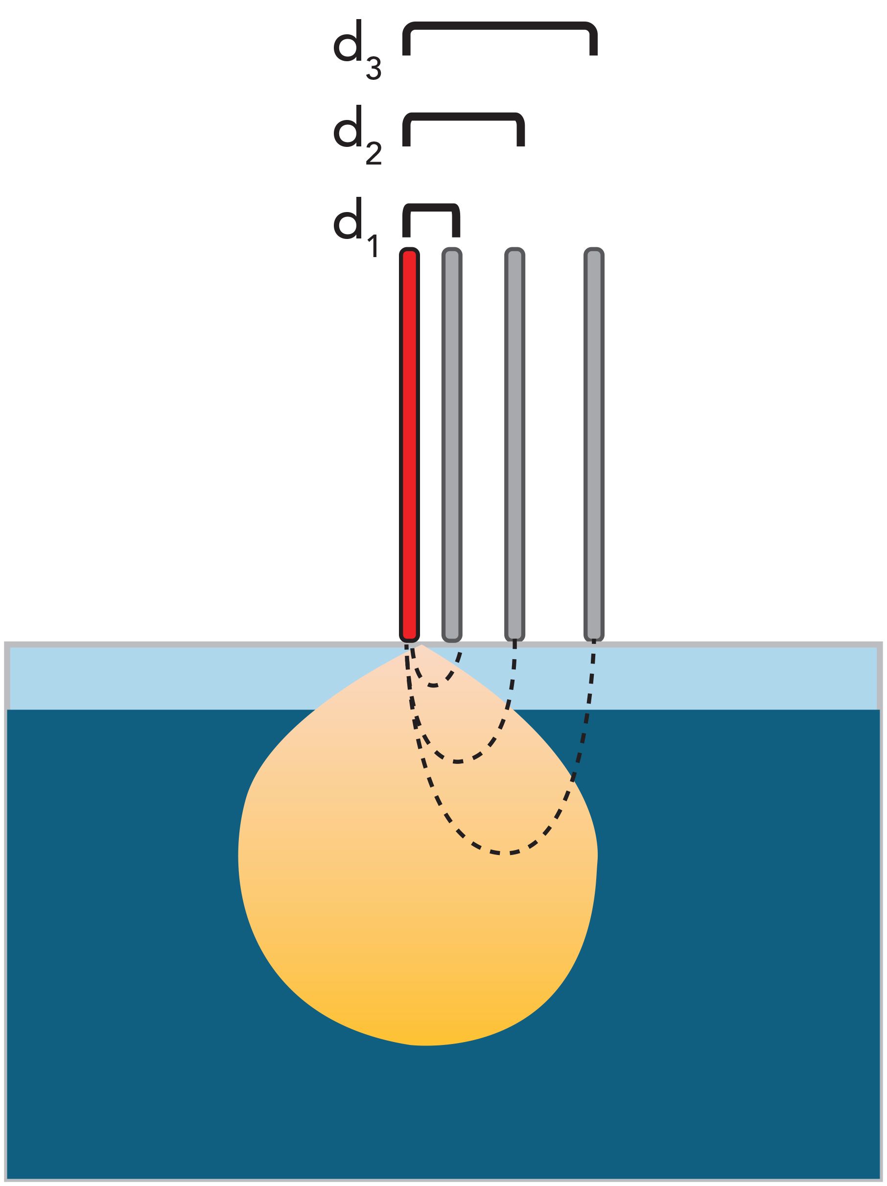
Understanding the effects of optical scattering on Raman signal recovery guides the design and selection of a fiber-optic probe. There are applications that should harness the effects of optical scattering to obtain a representative sample or minimize the effects of optical scattering from bubbles or particulates. Figure 2 shows the principle of photon scattering through a layered solid system and how fiber geometries can be tailored to optimize collection of a surface or subsurface Raman signal.
Figure 2: Understanding the principles of photon diffusion in turbid media can help with selection of a sampling probe. In turbid media, laser light delivered by the excitation fiber (shown in red) is scattered multiple times and results in a volume of scattered light (shown in yellow). Dashed lines show the path of surface-scattered and subsurface-scattered photons. Strategic placement of collection fibers (in grey) can optimize collection of Raman signal. In the case of a closely placed collection fiber, at distance d1, most of the collected signal comes from the surface. Longer collection fiber offsets from the excitation results in collection of signal from the surface with some subsurface signal, shown by d2, and mostly subsurface signal, shown by d3.

Figure 3 shows commercially available variants of sampling probes for Raman spectroscopy using optical fibers. A commonly used approach is backscattered Raman spectroscopy, which is highlighted in Figure 2. In backscattered Raman spectroscopy, incident and Raman-scattered light share the same optical path. This geometry allows the collection of a small volume. In the layered system shown in Figure 2, a backscattered probe will collect a signal from the surface layer. This type of probe geometry can be applied toward measurement in liquids, especially to reduce potentially deleterious effects of bubbles, turbidity in the liquid, or immiscible liquids. This type of probe can also be used to measure surfaces of solids.
Figure 3: An in-depth view of available fiber geometries for sampling probes, type of measurement provided, and their typical installations. Solid lines indicate delivery of the laser via an optical fiber, and defocused laser delivery indicated by a solid cylinder. Dashed lines indicate the position of collection fibers. There are many possibilities for offline measurements of solids or liquids, and a single analyzer with multiple “plug-and-play” probes can provide sampling flexibility. For inline or online measurements, it is important to consider the installation location in addition to the process’s optical scattering and chemical properties. (SORS = spatially offset Raman spectroscopy.)

To obtain signals from a larger volume, collection fibers can be offset from the excitation fiber. Large volumetric Raman spectroscopy uses a wide laser beam and multiple collection fibers to obtain data from surface and subsurface layers. Large volumetric sampling is a useful approach for in-process measurements of content uniformity or processing solids and turbid media. Spatially offset Raman spectroscopy (SORS) and transmission Raman spectroscopy utilize a single-point excitation with spatially offset collection fibers. In SORS, the fibers are typically in the same plane, and in transmission Raman spectroscopy, the collection fibers are typically 180° from the excitation fiber. These probes are used in solids applications ranging from through-container raw material identification to in-process quality assurance of processes involving solids or turbid media such as blending. Together, these four variants provide sampling flexibility from early development to manufacturing, in applications from laboratory quality assurance (QA) to in-process monitoring, testing, or control.
Analytical Model
There are many approaches to modeling spectroscopic data with either a univariate or multivariate model. Multivariate models are more commonly used, and these models can incorporate first principle bounds in the model (known as hard modeling) or be based on correlation or covariance without any a priori knowledge (known as soft modeling) (17). One important consideration for industrial applications is model transferability. Model transfer can refer to a) transfer across different instruments that measure the same process, or b) expanding the scope of an existing model to account for changes in process conditions, such as pressure, temperature, media composition, and reactor size. An excellent 2018 review on calibration transfer discusses how hardware design affects model transfer (18). Hardware built for high-accuracy with a uniform internal design, but with housing configurable to the installation environment, has a few advantages, including reproducible cross-instrument performance, compatibility with “turn-key” sampling probes, and ease of servicing. This flexible design allows the user to perform calibration transfer from instrument to instrument and prevents calibration rework in the event of subsampling, or a laser or detector change. Another important consideration is the choice of probe. Probe selection impacts representative sampling and model development, and certain probe optics may adversely affect model transferability or robustness in turbid media and solids (19,20).
Applications
Biopharmaceuticals
Raman spectroscopy has been successfully applied to upstream biopharmaceutical development in industrial settings since 2010 (17). Whether for cell culture or fermentation bioprocesses, in batch or continuous mode, Raman spectroscopy has many proven benefits, including the ability to simultaneously measure nutrients, metabolites, and cell viability. Raman spectroscopy also allows for the cross-scale method transfer from the laboratory to a current good manufacturing practice (cGMP) environment without significant method adjustments (21–23). Recent studies have demonstrated that Raman spectroscopy-based feedback control of glucose feeding or lactate accumulation in cell cultures has benefits of improved monoclonal antibody product quality and increased titer (24–26). Feedback-based control was also achieved in highly fluorescing cell culture and fermentation applications using wavelengths closer to shortwave infrared (1–1.4 μm) (27,28). More recent work has further expanded upstream monitoring to amino acids and titer predictions (29,30). In downstream applications, Raman spectroscopy can be equally powerful and we have seen recent successful cases of Raman spectroscopy used to quantify protein concentration and to monitor chromatography-based purification and crystallization (31,32).
Chemical Manufacturing
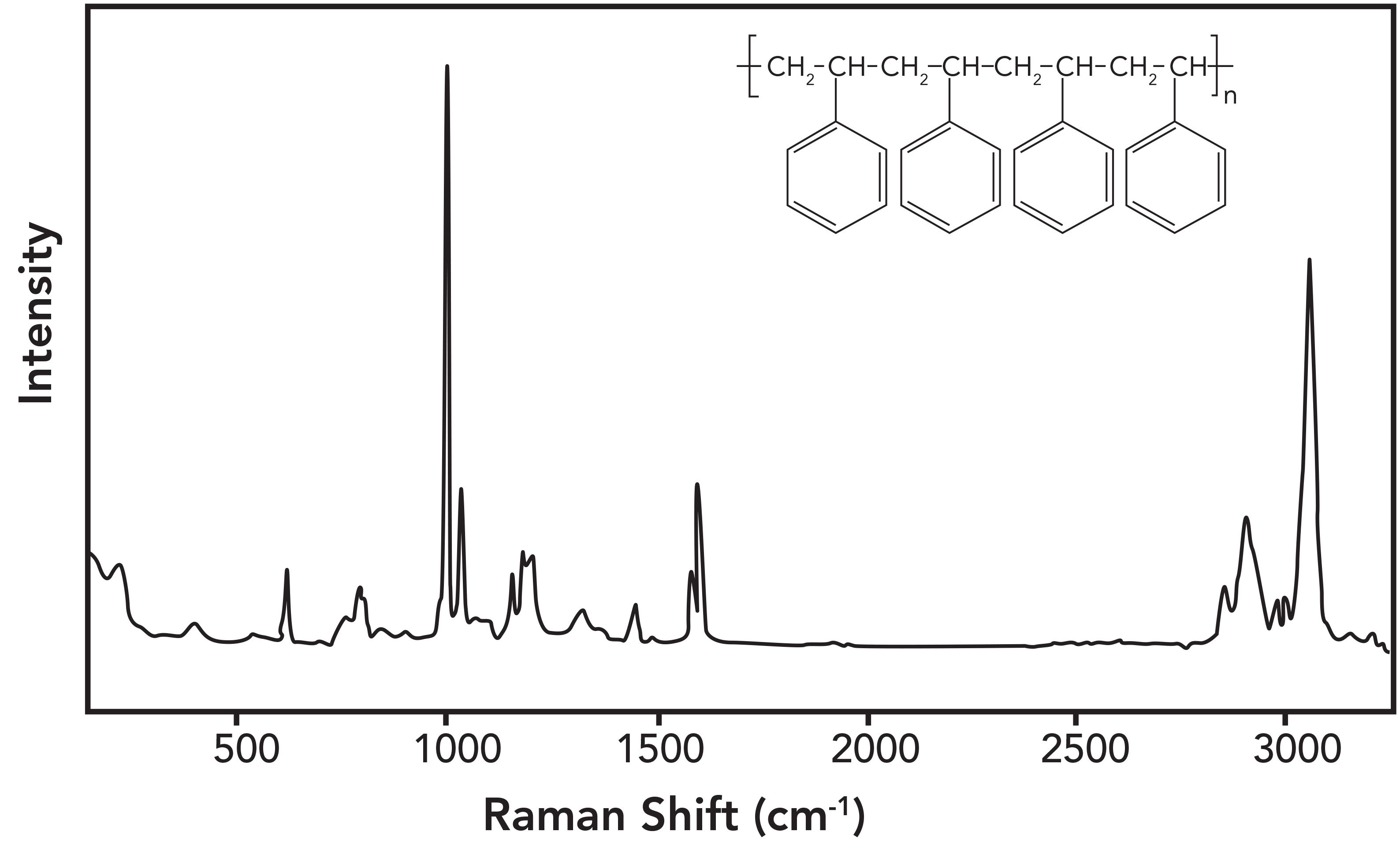
As performance demands on polymeric materials have increased, polymerization processes have become more complex and often require tighter control of process parameters. Enabling mega-volume processes that can consistently make a quality product safely have increased the demand for PAT with high uptime that enables real-time, closed-loop process control. An example of successful polymer analysis by Raman spectroscopy is polystyrene, an important polymeric material. Figure 4 shows a Raman spectrum of polystyrene. What should be noted about the spectrum is that the peaks correspond to known chemical moieties, and they are sharp, identifiable, and quantifiable, which makes robust process monitoring and control possible. Styrene polymerization can be easily quantified using the area or intensity ratio of the styrene vinyl bond at 1630 cm−1 to the 1000-cm-1 reference band.
Figure 4: The chemical structure of polystyrene (insert) and its Raman spectrum. The sharp Raman bands of polystyrene correspond to specific chemical structures and can be used to identify, monitor, and quantify the polymer.

Raman spectroscopy-derived concentration predictions closely match measurements carried out by gravimetry, but without the need for sample extraction. The work reported by Brun’s team indicates that Raman spectroscopy could be part of a control strategy to avoid process upsets and ensure quality of a polymer product (33). Specifically, these results demonstrate the utility of Raman spectroscopy for monitoring the polymerization of styrene into polystyrene. The data were able to be generated quickly (in a matter of seconds), and they corresponded well with data obtained by classical gravimetric methods that are known to give good results but are also cumbersome and time-consuming.
Csontos’s group developed an effective feedback control loop based on Raman spectroscopy for a hazardous exothermic oxidation reaction (34). They were able to determine the end point of the process reaction, following the quantification of reaction components and an unstable intermediate. This was another successful example of Raman spectroscopy giving visibility into an industrial chemical reaction allowing for real-time control of the overall process. Similarly, Hart’s group was able to develop an accurate Raman spectroscopy calibration model for end-point determination of an etherification, where the residual level of chloropyrazine starting material needed to be minimized (35). At the outset, they were aware the reaction may be scale dependent because a heterogenous base (K2CO3) was used. They successfully demonstrated that the calibration work for reaction end-point determination done at the laboratory scale was easily transferred to the scaled-up pilot plant, with predicted results closely matching manual off-line high-performance liquid chromatography results.
Raman spectroscopy has also been used to improve our understanding of the reaction kinetics of many processes, such as mechanochemical Knoevenagel condensations (36). In one study, crystallization properties were examined for the condensation of three fluorinated benzaldehyde derivatives and malononitrile by Hart’s group using in situ
Raman spectroscopy.
As we continue to find ways of dealing with climate change, Raman spectroscopy will play an increasingly significant role in controlling and optimizing industrial processes that involve greenhouse gases like CO2. Recently, Jinadasa’s group demonstrated the ability of Raman spectroscopy to effectively monitor speciation of a CO2 capture process for both the lean and rich amine streams (37). Other chemical processes involving carbon chemistry, such as the common Suzuki-coupling reactions of the C–C bond, can also benefit from Raman spectroscopy through yield optimization. In this scenario, parameters like temperature and concentration can be varied independently within reaction limits to find optimal operating conditions. In doing so, Heteni and Janagap were able to develop a robust partial least squares (PLS) model using their experimental Raman spectra, and online predictions from the model agreed well with offline gas chromatography–mass spectrometry (GC–MS) measurements of reaction yield (38).
Pharmaceuticals
Raman has been used to examine crystallization and the solid state form of small-molecule active pharmaceutical ingredients (API) since the 1980s (39). As in many industries, applications of Raman spectroscopy in small-molecule pharmaceuticals have surged in the past three decades as technology has improved. Because of these technological advances, Raman spectroscopy is now a practical analysis technique in the pharmaceutical laboratory and process environments. As a PAT in API manufacturing, Raman spectroscopy has demonstrated value from scientific understanding to process control. A recent paper by Nagy’s group highlights the industrial trend for using Raman spectroscopy for real-time monitoring and control for secondary processing batch steps (40). We are enthusiastic about recent reports in continuous solids monitoring and solid phase unit operations, including in situ control of crystallization and polymorphism, process-induced transformations, low-dose formulations, and tablet coating. Automated Raman spectroscopy measurements are compatible with the fast cycle times and can provide process understanding for continuous solids production approaches, including twin-screw granulation and hot melt extrusion (41,42).
Food and Beverage Industry
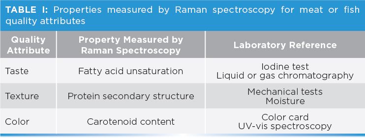
The food and beverage industry works in a highly regulated environment with strict controls on quality and safety. In this respect, there are similarities between food and beverage production and biopharmaceutics and small molecule API manufacturing. Although the PAT framework has been successfully used in pharmaceutical and biopharmaceutical manufacturing since 2004, its application has lagged in the food and beverage industry until recently. A more current interpretation of PAT in food and beverage is that it represents “a silent revolution in industrial quality control in food processing” (43). As a result, there are more reports of Raman spectroscopy in food processing (44,45). One application area that is particularly benefitting from Raman spectroscopy is meat and fish processing. Meat and fish quality is based on taste, texture, and appearance. All of these quality attributes have a basis in the specimen’s chemical properties, making Raman spectroscopy well-suited to providing a fast, non-destructive, and multiattribute quality measurement (46). Table I shows an overview of Raman spectroscopy-provided laboratory measurements for meat or fish quality attributes.

Laboratory studies demonstrate that Raman spectroscopy can provide both a fast and effective analytical method for determining iodine value, polyunsaturated fatty acids, monounsaturated fatty acids, and saturated fatty acids in samples of pork back fat (47–49). Even in a highly sophisticated meat processing plant, a fair amount of byproducts are generated. One strategy to minimize by-product waste is to use it as input for creating other valuable materials such as hydrolyzed proteins, and new research is showing the promise of Raman spectroscopy-based feedback control in this application (50,51).
Conclusions
Raman spectroscopy is a valuable PAT for many applications across multiple industries. The molecular specificity, ability to be directly coupled to a reaction vessel, and compatibility with solids, liquids, gases, and turbid media are advantages of Raman spectroscopy. Application success and rapid return-on-investment are realized in Raman spectroscopy-based feedback control because it enables in-process corrections and ensures process and product quality. The established history of process Raman spectroscopy in molecular identification, quantification, and process monitoring form the basis for continuing to expand Raman spectroscopy to new applications. We are enthusiastic about bringing Raman spectroscopy even further into industry through the adoption of industry 4.0 principles in PAT, increased automation, and even more integration of Raman spectroscopy into process control applications.
Acknowledgments
We thank Dr. Linda Kidder and Dr. Casey Kneale for helpful discussions during manuscript preparation.
References
- R.I. Lewis, H. Edwards, Handbook of Raman Spectroscopy: From the Research Laboratory to the Process Line (CRC Press, London, United Kingdom, 2001).
- N.L. Jestel, in Process Analytical Technology, K.A. Bakeev, Ed. (Blackwell Publishing, Oxford, United Kingdom, 2005).
- P. Vandenabeele, Practical Raman Spectroscopy: An Introduction (Wiley, New York, New York, 2013).
- G. Turrell, J. Corset, Raman Microscopy Developments and Applications (Elsevier, New York, New York, 1996).
- M. Delhaye, P. Dhamelincourt,J. Raman Spectrosc. 3(1), 33–43 (1975).
- M.D. Morris, G.S. Mandair, in Raman, Infrared, and Near-Infrared Chemical Imaging, Y.O. Slobodan Šašić, Ed. (John Wiley & Sons, Inc., New York, New York, 2010), pp. 109–131.
- S. Nakashima, J. Phys. Condens. Matter 16(2), S25–S37 (2004).
- A. Zoubir, Ed. Raman Imaging: Techniques and Applications (Springer Berlin Heidelberg, Berlin, Heidelberg, Germany, 2012), vol. 168. https://doi.org/10.1007/978-3-642-28252-2.
- R.M. Stöckle, Y.D. Suh, V. Deckert, R. Zenobi, Chem. Phys. Lett. 318 (1), 131–136 (2000). https://doi.org/10.1016/S0009-2614(99)01451-7.
- E. Bailo, and V. Deckert, Chem. Soc. Rev. 37(5), 921–930 (2008). https://doi.org/10.1039/B705967C.
- P.L. Stiles, J.A. Dieringer, N.C. Shah, and R.P. Van Duyne, Annu. Rev. Anal. Chhttps://doi.org/10.1007/978-3-642-28252-2em. 1(1), 601–626 (2008). https://doi.org/10.1146/annurev.anchem.1.031207.112814.
- C.L. Haynes, A.D. McFarland, and R.P. Van Duyne, Anal. Chem. 77, 338A–346A (2005).
- J.X. Cheng and X.S. Xie, J. Phys. Chem. B. 108(3), 827–840 (2004). https://doi.org/10.1021/jp035693v.
- G.L. Eesley, Coherent Raman Spectroscopy (Elsevier, New York, New York, 2013).
- G.J. Gervasio, M.J. Pelletier, J. Process Anal. Chem. 3(1/2), 7–11 (1997).
- E. Berrocal, D.L. Sedarsky, M.E. Paciaroni, I.V. Meglinski, and M.A. Linne, Opt. Express 15(17), 10649–10665 (2007). https://doi.org/10.1364/OE.15.010649.
- K.A. Esmonde-White, M. Cuellar, C. Uerpmann, B. Lenain, and I.R. Lewis, Anal. Bioanal. Chem. 409(3), 637–649 (2017). https://doi.org/10.1007/s00216-016-9824-1.
- J.J. Workman, Appl. Spectrosc. 72(3), 340–365 (2018). https://doi.org/10.1177/0003702817736064.
- E.F. Olsen, C. Baustad, B. Egelandsdal, E.O. Rukke, and T. Isaksson, Meat Sci. 85(1), 1–6 (2010). https://doi.org/10.1016/j.meatsci.2009.12.008.
- H.J. van Manen, R. Bloemenkamp, and O.F. van den Brink, Appl. Spectrosc. 63(3), 378–380 (2009).
- B. Berry, J. Moretto, T. Matthews, J. Smelko, K. Wiltberger, Biotechnol. Prog. 31(2), 566–577 (2015). https://doi.org/10.1002/btpr.2035.
- H. Mehdizadeh, D. Lauri, M.K. Karry, M. Moshgbar, R. Procopio-Melino, and D. Drapeau, Biotechnol. Prog. 31(4), 1004–1013 (2015). https://doi.org/10.1002/btpr.2079.
- N.R. Abu-Absi, B.M. Kenty, M.E. Cuellar, M.C. Borys, S. Sakhamuri, D.J. Strachan, M.C. Hausladen, and Z.J. Li, Biotechnol. Bioeng. 108(5), 1215–1221 (2011). https://doi.org/10.1002/bit.23023.
- S. Craven, J. Whelan, and B. Glennon, J. Process Control 24(4), 344–357 (2014). https://doi.org/10.1016/j.jprocont.2014.02.007.
- B.N. Berry, T.M. Dobrowsky, R.C. Timson, R. Kshirsagar, T. Ryll, and K. Wiltberger, Biotechnol. Prog. 2016, 32(1), 224–234 (2016). https://doi.org/10.1002/btpr.2205.
- T.E. Matthews, B.N. Berry, J. Smelko, J. Moretto, B. Moore, and K. Wiltberger, Biotechnol. Bioeng. 113(11), 2416–2424 (2016). https://doi.org/10.1002/bit.26018https://doi.org/10.1002/bit.26018.
- T.E. Matthews, J.P. Smelko, B. Berry, S. Romero-Torres, D. Hill, R. Kshirsagar, and K. Wiltberger, Biotechnol. Prog. 34(6), 1574–1580 (2018). https://doi.org/10.1002/btpr.2711.
- S. Goldrick, D. Lovett, G. Montague, and B. Lennox, Bioengineering 5(4), 79 (2018). https://doi.org/10.3390/bioengineering5040079.
- H. Bhatia, H. Mehdizadeh, D. Drapeau, S. Yoon, Eng. Life Sci. 18(1), 55–61 (2018). https://doi.org/10.1002/elsc.201700084.
- S. André, L.S. Cristau, S. Gaillard, O. Devos, E. Calvosa, and L. Duponchel, Anal. Chim. Acta. 892, 148–152 (2015). https://doi.org/10.1016/j.aca.2015.08.050.
- D. Yilmaz, H. Mehdizadeh, D. Navarro, A. Shehzad, M. O’Connor, and P. McCormick, Biotechnol. Prog. 36(3), e2947 (2020). https://doi.org/10.1002/btpr.2947.
- F. Feidl, S. Garbellini, F.M. Luna,S. Vogg, J. Souquet, H. Broly, M. Morbidelli, and A. Butté, Processes 7(10), 683 (2019). https://doi.org/10.3390/pr7100683.
- N. Brun, I. Youssef, M.C. Chevrel, D. Chapron, C. Schrauwen, S. Hoppe, P. Bourson, and A. Durand, J. Raman Spectrosc. 44(6), 909–915 (2013). https://doi.org/10.1002/jrs.4279.
- I. Csontos, H. Pataki, A. Farkas, H. Bata, B. Vajna, Z.K. Nagy, G. Keglevich, G.J. Marosi, Org. Process Res. Dev. 19(1), 189–195 (2015). https://doi.org/10.1021/op500015d.
- R.J. Hart, N.I. Pedge, A.R. Steven, and K. Sutcliffe, Org. Process Res. Dev. 19(1), 196–202 (2015). https://doi.org/10.1021/op500027w.
- S. Haferkamp, W. Kraus, and F. Emmerling, J. Mater. Sci. 53(19), 13713–13718 (2018). https://doi.org/10.1007/s10853-018-2492-0.
- M.H.W.N. Jinadasa, K.J. Jens, L.E. Øi, and M. Halstensen, Energy Procedia 114, 1179–1194 (2017). https://doi.org/10.1016/j.egypro.2017.03.1282.
- D. Hetemi and S. Janagap, Vib. Spectrosc. 100, 93–98 (2019). https://doi.org/10.1016/j.vibspec.2018.11.008.
- B.A. Bolton, P.N. Prasad, J. Pharm. Sci. 70(7), 789–793 (1981). https://doi.org/10.1002/jps.2600700720.
- B. Nagy, A. Farkas, E. Borbás, P. Vass, Z.K. Nagy, and G. Marosi, AAPS PharmSciTech 20(1), 1 (2019). https://doi.org/10.1208/s12249-018-1201-2.
- W. Meng, A. Román, S. Panikar, C. O’Callaghan, S. Gilliam, R. Ramachandran, and F. Muzzio, Adv. Powder Technol. 30 (2019). https://doi.org/10.1016/j.apt.2019.01.017.
- L. Arnfast, J. van Renterghem, J. Aho, J. Bøtker, D. Raijada, S. Baldursdóttir, T. De Beer, and J. Rantanen, Pharmaceutics 12(2), 116 (2020). https://doi.org/10.3390/pharmaceutics12020116.
- F. van den Berg, C.B. Lyndgaard, K.M. Sørensen, and S.B. Engelsen, Trends Food Sci. Technol. 31(1), 27–35 (2013). https://doi.org/10.1016/j.tifs.2012.04.007.
- H. Jin, Q. Lu, X. Chen, H. Ding, H. Gao, and S. Jin, Appl. Spectrosc. Rev. 51(1), 12–22 (2016). https://doi.org/10.1080/05704928.2015.1087404.
- D. Yang and Y. Ying, Appl. Spectrosc. Rev. 46(7), 539–560 (2011). https://doi.org/10.1080/05704928.2011.593216.
- A.M. Herrero, Food Chem. 107(4), 1642–1651 (2008). https://doi.org/10.1016/j.foodchem.2007.10.014.
- E.F. Olsen, E.O. Rukke, A. Flåtten, and T. Isaksson, Meat Sci. 76(4), 628–634 (2007). https://doi.org/10.1016/j.meatsci.2007.02.004.
- D.T. Berhe, C.E. Eskildsen, R. Lametsch, M.S. Hviid, F. van den Berg, and S.B. Engelsen, Meat Sci. 111, 18–26 (2016). https://doi.org/10.1016/j.meatsci.2015.08.009.
- M. Nache, R. Scheier, H. Schmidt, and B. Hitzmann, Chemom. Intell. Lab. Syst. 142, 197–205 (2015). https://doi.org/10.1016/j.chemolab.2015.02.002.
- S.G. Wubshet, J.P. Wold, N.K. Afseth, U. Böcker, D. Lindberg, F.N. Ihunegbo, and I. Måge, Food Bioprocess Technol. 11(11), 2032–2043 (2018). https://doi.org/10.1007/s11947-018-2161-y.
- S.G. Wubshet, J.P. Wold, U. Böcker, K.W. Sanden, and N.K. Afseth, Food Control 95, 267–273 (2019). https://doi.org/10.1016/j.foodcont.2018.08.017.
Karen Esmonde-White, Michael Kester, Maryann Cuellar, and Ian Lewis are with Kaiser Optical Systems, Inc., an Endress + Hauser company.Direct correspondence to: karen.esmonde-white@endress.com

AI-Powered SERS Spectroscopy Breakthrough Boosts Safety of Medicinal Food Products
April 16th 2025A new deep learning-enhanced spectroscopic platform—SERSome—developed by researchers in China and Finland, identifies medicinal and edible homologs (MEHs) with 98% accuracy. This innovation could revolutionize safety and quality control in the growing MEH market.
New Raman Spectroscopy Method Enhances Real-Time Monitoring Across Fermentation Processes
April 15th 2025Researchers at Delft University of Technology have developed a novel method using single compound spectra to enhance the transferability and accuracy of Raman spectroscopy models for real-time fermentation monitoring.
Nanometer-Scale Studies Using Tip Enhanced Raman Spectroscopy
February 8th 2013Volker Deckert, the winner of the 2013 Charles Mann Award, is advancing the use of tip enhanced Raman spectroscopy (TERS) to push the lateral resolution of vibrational spectroscopy well below the Abbe limit, to achieve single-molecule sensitivity. Because the tip can be moved with sub-nanometer precision, structural information with unmatched spatial resolution can be achieved without the need of specific labels.