New Approach for Optimizing a Monoclonal Antibody Biotherapeutic in Different Formulations
A prerequisite for a successful biotherapeutic formulation is one where the protein is stable and correctly folded.
David Gregson and Lindsay Cole, Applied Photophysics Limited
A prerequisite for a successful biotherapeutic formulation is one where the protein is stable and correctly folded. The new technique of dynamic multi-mode spectroscopy (DMS) was used to study the stability of a monoclonal antibody biotherapeutic formulated in acetate and lactate buffers. The samples were measured several times over a period of weeks and it became apparent that the antibody behaved differently as it aged in the two formulations, with the lactate formulation imparting greater robustness than the acetate.
Experimental
DMS uses two or more spectroscopic probes to generate full near- or far-UV spectra of a protein as a function of continuously changing temperature, which provides both structural and thermodynamic information in a single, sample-efficient experiment. In this study, each DMS dataset was obtained in under 100 min and used only 65 μg of protein. Subsequent analyses of the data were carried out using a proprietary global analysis program, an integral part of the DMS technique.
Results and Discussion
Robust Tm values were calculated from the CD temperature profiles by global analysis. Significant differences in the mid-points of the first and third transitions were observed; the mid-points of the second transitions were the same within experimental error. The first transition was lower in the lactate than in the acetate; the third transition was the most significant and best defined in both acetate and lactate buffers and was higher in the lactate formulation by 1.8 °C. There was significant variation in the individually calculated enthalpies, an inevitable consequence of the degree of overlap of transitions in these complex systems.
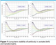
The CD spectra of the DMS data were used to identify changes in the secondary structure of the antibody. Both acetate and lactate formulations maintain the antibody in its mainly β-sheet conformation as they age and, initially, the conformation changes on heating are very similar, with each formulation taking a virtually identical structural pathway to a recognizable unfolded state (Figure 1, top). As the samples age, the denaturation pathway in acetate changes dramatically but remains virtually identical in lactate (Figure 1, bottom), suggesting greater stability.

Figure 1
The absorption spectra of the DMS data were used to identify the onset of aggregation. The temperature of onset of aggregation in acetate was found to decrease significantly with sample age, whereas no tendency to aggregate was found for the lactate irrespective of the age of the sample.
Conclusions
The results suggest that the antibody is more stable in the lactate than in the acetate formulation despite its significantly lower Tm1, which if taken in isolation might suggest the contrary. It is interesting to note that the data presented here support opinion voiced at a recent biocalorimetry conference (1), that Tm on its own may not be a particularly good indicator of stability and that resistance to aggregation once unfolding has occurred is likely to be a better indicator. The ability of DMS to measure structural, thermodynamic, and aggregation behavior in a single experiment highlights the value of using the technique in formulation testing during biotherapeutic antibody development.
References
(1) Applications of Biocalorimetry, Heidelberg, July 7–10, 2008.

Applied Photophysics Limited
201-205 Kingston Road
Leatherhead, Surrey
United Kingdom
Tel. +44 1372 386537
Fax +44 1372 386477
E-mail: sales@photophysics.com
Website: www.photophysics.com
