SERS Comes of Age for Molecular Detection
Special Issues
Since it was first described in 1974, surface-enhanced Raman spectrometry (SERS) has been thought to offer significant potential for a range of different applications. The theoretical sensitivity and specificity envisaged for this powerful technique has engaged scientists for many years, but practical challenges have hindered its routine adoption. Now, a new approach combines a robust and reliable substrate with expertise in surface chemistry and molecular biology on a platform that can be adapted for a wide variety of Raman instrumentation and customized routine applications.
This article illustrates the latest developments in the field of ultrarapid and highly multiplexed molecular detection at trace level. Data from labeled oligonucleotides show how the sensitivity, specificity, and mass screening capability of the substrate can be customized by combining the patterned surface-enhanced Raman spectrometry (SERS) substrate with application-specific chemistry.
The demand for rapid molecule recognition at trace level is required in many application areas, including molecular diagnostics, theranostics, chemical analysis, and chemical and biological threat detection. Many workers have presented SERS technology as having the potential to revolutionize molecular detection. However, to be accepted as an industry standard, it is necessary for any novel measurement method to demonstrate a strict set of criteria, common to all branches of science.
These include
- High sensitivity
- Exquisite specificity
- Trace-level detection capabilities from low sample volumes
- Fast analysis
- Potential for automation and high throughput
- User-friendly assay formats
- Reliable robust data
Timely Development
The first commercially available SERS-active surface (Klarite, D3 Technologies Ltd, Glasgow, UK) was introduced in 2005. It has been shown to deliver robust, reproducible results (1–3). Since then, there has been a surge in advancement (Figure 1). Early in 2008, rapid mass screening at trace levels using SERS surfaces was demonstrated (4). Important for a wide range of applications, this development brings SERS to the stage, where it answers the demands laid down for routine detection systems.
The combination of SERS-active substrate, surface chemistry, and molecular biology expertise with the Raman instrumentation now available provides the features and performance required by the marketplace. SERS looks set to come of age.

Figure 1
Adding Functionality with Surface Chemistry
By combining a patterned SERS-active substrate with surface chemistry (Figure 2), SERS surfaces can be functionalized to produce high binding specificity. The result is quantitative, simultaneous multianalyte detection.

Figure 2
With controlled chemistry, surface-enhanced resonance Raman scattering (SERRS) can be performed at a nanostructured surface. This technique is suitable for a varied range of applications in biodiagnostics, including gene probes and DNA detection. Recent studies have shown reproducible SERRS spectra from dye-labeled oligonucleotides (4).
Comparison of the fluorescence and Raman spectra of a commercially available dye (Figure 3) highlights the higher discrimination power of SERRS compared to fluorescence. The narrow lines of the Raman spectrum allow unambiguous signal recognition in a highly multiplexed configuration and with instrument platforms using single-excitation wavelength and readout optics.
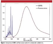
Figure 3
Adding customized surface chemistry to highly reproducible substrates provides other fundamental advantages. For example, a major cause of poor reproducibility in SERS measurements is a lack of consistency in how sampling is done and how target molecules are delivered to the surface. Ensuring a good analyte-to-substrate attachment is extremely important in guaranteeing uniform and reproducible sampling. This results in more reliable, reproducible, and quantitative results — solving one of the challenges that has hindered the adoption of SERS as a routine analytical technique.
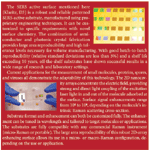
The flexibility of chemistry and substrate engineering means that the Klarite platform lends itself to full customization. Both chemistry and substrate can be tailored specifically for a particular application.
Multiplexed DNA Measurement
An example of the versatility and potential of combining SERS substrates with customized chemistry is given in Figures 3 and 4. Oligonucleotides were modified at one end with a group designed to adhere to the surface and at the other with different fluorophores. They were applied to 4 mm × 4 mm Klarite surface in a simple matrix pattern (Figure 4a).
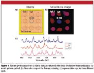
Figure 4
The scanning of the whole surface, comprising over 4,000,000 substrate nanowells, was recorded by a high-performance Raman spectrometer (StreamLine, Renishaw plc, Wotton-under-Edge, UK) in less than 3 min using low power density and low numerical aperture optics.
In-situ identification of three analytes is illustrated in Figures 4b and 4c.
The image in Figure 4b illustrates the huge amount of information that can be generated by the combination of the Klarite SERS platform with Raman imaging. Spatial resolution and multiple analyte identification can be obtained in a highly multiplexed configuration. Only very small sample volumes are required, and limits of detection can be at femtomole to attomole concentrations.
Raman "fingerprinting" also allows in-situ molecular recognition in this highly multiplexed configuration (Figure 5). Importantly, information about individual molecules within a group of molecules on the matrix can be seen easily.
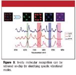
Figure 5
Conclusion
In-situ molecular detection, identification, and quantitation in a multiplexed configuration are achievable with SERS. Building on the classic advantages of Raman, SERS technology has demonstrated the features and performance required to firmly establish it as a technology for future diagnostic applications.
Professor Ewen Smith is CEO and Dr. Caterina Netti is Director of Technology at D3 Technologies Ltd., Glasgow, UK. Robert Stokes is a Research Fellow at the Centre for Molecular Nanometrology of the University of Strathclyde, UK.
References
(1) T. Alexander, Anal. Chem. 80, 2817–2825 (2008).
(2) G.S. Mandair, K.A. Dehring, B.J. Roessler, and M.D. Morris, Proc. SPIE 6093 115–121 (2006).
(3) A. Szeghalmi, S. Kaminskyj, P. Roesch, J. Popp, and K.M. Gough, J. Phys. Chem. B 111, 12916–12924 (2007).
(4) R. Stokes et al., Chem. Commun. 2811–2813 (2007).

AI-Powered SERS Spectroscopy Breakthrough Boosts Safety of Medicinal Food Products
April 16th 2025A new deep learning-enhanced spectroscopic platform—SERSome—developed by researchers in China and Finland, identifies medicinal and edible homologs (MEHs) with 98% accuracy. This innovation could revolutionize safety and quality control in the growing MEH market.
New Raman Spectroscopy Method Enhances Real-Time Monitoring Across Fermentation Processes
April 15th 2025Researchers at Delft University of Technology have developed a novel method using single compound spectra to enhance the transferability and accuracy of Raman spectroscopy models for real-time fermentation monitoring.
Nanometer-Scale Studies Using Tip Enhanced Raman Spectroscopy
February 8th 2013Volker Deckert, the winner of the 2013 Charles Mann Award, is advancing the use of tip enhanced Raman spectroscopy (TERS) to push the lateral resolution of vibrational spectroscopy well below the Abbe limit, to achieve single-molecule sensitivity. Because the tip can be moved with sub-nanometer precision, structural information with unmatched spatial resolution can be achieved without the need of specific labels.