In Situ FTIR Analysis of Soils for Forensic Applications
Special Issues
A huge amount of information is contained in the FTIR spectra of soils in the mid infrared (MIR) region (4000 to 400 cm-1). The spectra provide an overall chemical profile of the soil, encompassing fundamental vibrations of both the organic and mineral components. Interpretation of the spectrum of individual soils can provide a powerful means of differentiating between samples and therefore has considerable potential for use in forensic applications, and indeed we have successfully used laboratory-based FTIR analysis of soil to provide evidence in forensic casework. In recent years handheld FTIR spectrometers have become available and this makes it possible for in situ or field-based FTIR analysis of soils at a crime scene. However, reliable and tested protocols are not yet available for field-based FTIR analysis of soil. This paper discusses the sampling options for field-based FTIR of soil and describes tests of the methodology we are developing, for a handheld FTIR, on soil samples tested in the context of a mock crime scene.
A huge amount of information is contained in the Fourier transform infrared (FT-IR) spectra of soils in the mid-infrared region (4000–400 cm-1). The spectra provide an overall chemical profile of the soil, encompassing fundamental vibrations of both the organic and mineral components. Interpretation of the spectrum of individual soils can provide a powerful means of differentiating between samples and therefore has considerable potential for use in forensic applications, and indeed we have successfully used laboratory-based FT-IR analysis of soil to provide evidence in forensic casework. In recent years handheld FT-IR spectrometers have become available, and they enable in situ or field-based FT-IR analysis of soils at a crime scene. However, reliable and tested protocols are not yet available for field-based FT-IR analysis of soil. This article discusses the sampling options for field-based FT-IR of soil and describes tests of the methodology we are developing, for a handheld FT-IR instrument, to test soil samples in the context of a mock crime scene.
Historically, soil has not always been considered in a forensic context, but in recent years there has been a considerable increase in interest in forensic soil science (1,2). Soil can potentially provide both important evidence and also intelligence in forensic cases, and despite being present on articles such as footwear, spades, and vehicles it has often been overlooked; soil can help link suspects to a crime scene or provide geological information that could narrow down the geographical location where the crime was committed. When considering possible methods for characterizing soil for forensic applications, Fourier transform infrared (FT-IR) spectroscopy is a method that has many advantages. FT-IR allows characterization of very small samples of material, is nondestructive, requires little sample preparation, and gives an overall chemical profile that encompasses both organic and inorganic constituents. This is highly advantageous for soil because it means that from a single IR spectrum, aspects of the mineralogy and the nature of the soil organic matter present can be determined. In addition, the presence of any contaminants can be detected. With careful sampling and sample preparation, relative proportions of minerals and organic matter can also be established. IR spectra can therefore be used for comparison of and discrimination between soil samples.
IR Spectra of Soil
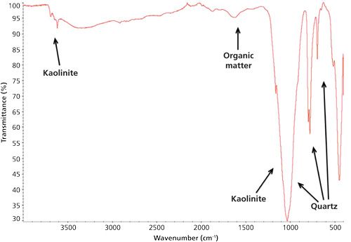
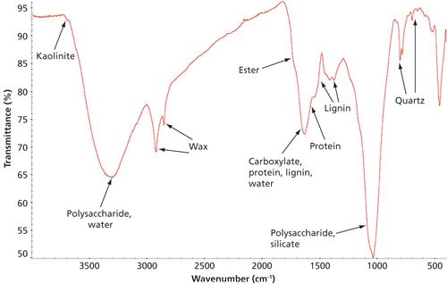
Soil is a highly complex and variable material (3) and can range from a mineral soil with almost no organic matter present to an entirely organic soil or peat. This is reflected in the very different IR spectra soil produces in the mid-IR region (4000–400 cm-1) because of the fundamental vibrations of the components present (see Figures 1 and 2). The minerals present in soil are predominantly silicates or carbonates but can also include other types of minerals such as sulfates. Clay minerals (aluminosilicates) play an important part in the properties of a soil (4,5) and have OH groups present with free OH groups (no OH bonding) and give very sharp well-defined absorption bands in the OH stretching region, often at higher frequencies than the organic OH (approximately 3700–3500 cm-1). These bands are highly diagnostic and, combined with bending vibrations, can allow the nature of the clay minerals present to be established. The bands arising from stretching vibrations are strong bands close to 1000 cm-1 for silicates, and close to 1400 cm-1 for carbonates. Generally, in soil these stretching vibrations arise from a combination of minerals and are not usually very diagnostic of the specific minerals present, but the pattern is representative of the soil and can be useful for discrimination. Bending vibrations (below 900 cm-1) can be used to identify the minerals and, for example, differentiate between calcite (calcium carbonate), aragonite (a different form of calcium carbonate), and dolomite (a calcium magnesium carbonate) (6,7). Silicate minerals can also be identified from their deformations, for example, quartz can be readily identified by a sharp doublet at 797 and 779 cm-1.
Organic material originates from vegetation and so the spectral features have much in common with spectral features of vegetation, although as it degrades it leads to the formation of other organic compounds such as humic and fulvic acids. The broad OH stretch between approximately 3600 and 3200 cm-1 is characteristic of soil organic matter (SOM), and the C-O stretch at 1030 cm-1 is characteristic for polysaccharide and clashes with the silicate stretch if minerals are present (8). The CH stretching region can show very sharp CH2 stretches at 2920 and 2850 cm-1 if long-chain molecules are present or more of a broad absorption if the material is more polysaccharide based. Other bands can be identified for organic matter, including ester peaks, carboxylic acid peaks and amide (protein) peaks.
Most soils will have a combination of mineral and organic matter, so features of both will appear in their IR spectrum. This is useful for forensic applications because it can allow discrimination even when working in areas with a similar underlying geology. It also means that if both organic matter and mineralogy of two samples match closely it makes it very much more likely that the samples are related, as they are effectively independent components. Any contaminants present in the soil can also provide a further element for comparison and strengthen the likelihood that two samples are related. For example, if you were comparing soil from a footprint found at a crime scene to soil from a suspect’s footwear, and the person wearing the footwear had crossed a garden and then a crumbling patio, carbonate from the patio might appear in the soil samples. If the footwear and crime scene soil samples showed IR spectra with very similar silicate profiles and SOM profiles with the presence of carbonate as a contaminant, then it could be judged highly unlikely that the combined profiles were obtained by coincidence.
It should however be noted that IR spectral patterns are very dependent on careful soil sampling, sample processing, and sampling method for FT-IR. Care must be taken to ensure reliable and reproducible spectra are obtained to make a valid comparison.
In Situ FT-IR Analysis of Soil
When carrying out IR analysis of forensic soil samples in the laboratory the samples can be dried and milled, and if sufficient soil is available a protocol for sample preparation (developed as part of the EPSRC SoilFit project) (9) can be followed (10). Sampling options for FT-IR of soils in the laboratory include transmission, in which KBr disks need to be prepared from the milled soil. Historically, this was the method used for IR analysis of soil, but it is very time consuming and requires an adjustment of the concentration of the sample to ensure that the IR spectrum is of a suitable intensity. This sampling method is not an option for in situ FT-IR analysis of soil, but methods such as diffuse reflectance and diamond attenuated total reflectance (DATR) can be used in the field. However, until recently, a bigger issue in terms of attempting in situ FT-IR analysis of soils was the lack of development of handheld FT-IR spectrometers. However, handheld FT-IR instruments have become available, which opens up the possibilities for field-based measurements. DATR can be regarded as the modern sampling alternative to transmission spectra, and it is described as “transmission like” because the spectra are similar in appearance to transmission spectra (7). A DATR sampling accessory has the advantages of allowing spectra of unprocessed soil to be recorded, automatically producing spectra of the right concentration, and being nondestructive. Spectra can also be recorded of very small samples, which can be an important consideration for forensic work. ATR does require intimate contact between the sample and the sampling window, but this contact can be easily achieved by smearing a sample on the window or by applying pressure through a torque-limited pressure arm.
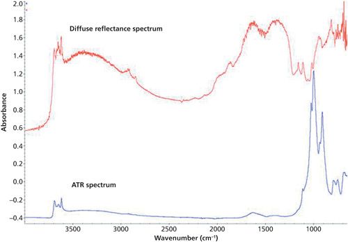
An alternative sampling option for FT-IR that is also suitable for in situ applications is diffuse reflectance or diffuse reflectance infrared Fourier transform spectroscopy (DRIFTS). In this case, the IR radiation is reflected from the external and internal surfaces of the sample; this approach is also nondestructive and requires little sample preparation. Diffuse reflectance is generally the accepted sampling method for soil spectroscopy in the near-infrared (NIR) region (11), and for this reason it often appears to be the preferred method in the mid-IR region. However, diffuse reflectance spectra in the mid-IR region have a very different appearance to ATR or transmission spectra-unless the samples are diluted with KBr, which is unlikely to be done in the field; the neat soil has too strong an absorbance, which leads to inversion in some areas of the spectrum (for example, silicate or carbonate) and gives rise to artifactual peaks (12). In the case of peat, DRIFTS on neat samples may not necessarily lead to inversion, but can lead to a lack of variation in intensity of many bands. In the mid-IR region the spectra arise from fundamental vibrations, rather than from the overtones and combinations of the NIR region, so it is easier to interpret the spectra and determine what components are present. However, interpretation can be a bit harder for the DRIFTS spectra compared to the DATR spectra. In Figure 3 there is an illustration of the same soil sampled by DRIFTS and DATR. There may be some advantages to having the stronger overtones that are seen in the diffuse spectra, and the larger sampling area. In situ analysis is on unmilled soil in the fresh state, so in addition to particle size variation and inhomogeneity there are also moisture issues. Generally, for ATR of unmilled spectra (see Figure 3) there is a more sloping baseline. There is also lower intensity of most mineral bands but greater intensity of clay minerals and SOM, which can have advantages. Currently, there are no methods or robust protocols for the in situ FT-IR analysis of soils and we are working to develop suitable methodology that will allow soil analysis in the field for a range of purposes including soil monitoring, agricultural, and forensic work.
In Situ FT-IR Analysis of Soil at a Mock Crime Scene
A mock crime scene, set up as part of the MiSAFE project, provided a useful means of testing the methodology we are developing for in situ FT-IR analysis of soil (13,14).
Materials and Methods
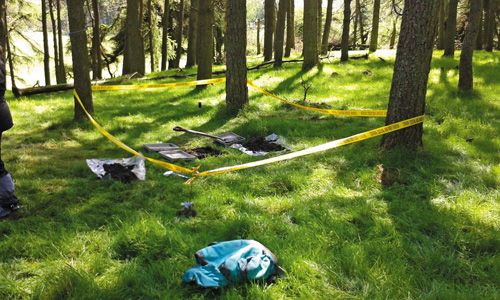
At a simulated crime scene a packet of drugs was buried, and the spade used was collected and stored as if concealed by the perpetrators. One week later, the crime scene (see Figure 4) was visited and control soil samples were taken from the topsoil and subsoil at the site of the buried drugs. In addition, soil samples were taken from a context comparison site 30 m away, an alibi site identified by a suspect, and a range of other comparison sites in the near vicinity (improved grassland pasture, unimproved grassland pasture, and an arable field).
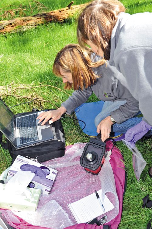
FT-IR analysis was carried out in situ on the four replicates of the topsoil samples taken from the crime scene (see Figure 5), and subsequently identical methodology was used to analyze the remaining field condition (field wet and unmilled) soil samples in the laboratory. The FT-IR spectra were recorded on an Agilent 4100 Exoscan handheld FT-IR spectrometer using a DATR sampling system in the 4000-650 cm-1 range, with 64 scans at a resolution of 4 cm-1. The methodology involved applying a thin, wet smear of soil, using a clean gloved finger. The smear of soil was then allowed to dry for several seconds before the spectrum was recorded. If the IR spectrum of the soil recorded after this period of time remained dominated by water, then an additional drying period was allowed and the IR spectrum was re-recorded. This was repeated again as necessary.
Results
For some of the samples (particularly the topsoil and subsoil from the crime scene) it was possible to smear a very thin layer of the soil onto the DATR sampling window that dried quickly and produced IR spectra with relatively low moisture and noise levels. For other samples (in particular the arable, improved grassland, and alibi sites) it was harder to get a thin layer of soil on the sampling window, and consequently drying times were longer (up to 2 min). Even after allowing a longer drying period, for some samples, a greater proportion of liquid water was retained and appeared in the IR spectra. However, the presence of water did not obscure all the relevant peaks that were features of the soil. In some spectra, appreciable amounts of water vapor appeared, mostly as the sample was drying. In general, the spectra had relatively low noise levels despite the fact that the samples were unmilled and of variable particle size. The unmilled condition of the soils did result in some of the mineral peaks being less well resolved and the baseline having a variation in slope. However, in the forensic context this lower resolution and baseline slope variation can be regarded as discriminatory for soils rather than posing a problem. Generally, replication of the IR spectra for each different soil that was sampled was good given the very small sample size (the DATR only requires a 2-µm-thick layer on a 1-mm-diameter sampling window), the field condition of the soils, and the likely natural heterogeneity.
The IR spectra of the soil samples had features from both the SOM and minerals present, which could be used in the comparisons between them. Although the mineral peaks in the spectra were not all well resolved, distinct clay minerals peaks were present, with kaolinite (3695 and 3620 cm-1) detectable in most of the samples and strongest in the subsoil sample from the crime scene. There was also evidence for illite or mixed layer clay in the context sample. Despite limited resolution of some of the mineral peaks, a similar pattern of bands in the main silicate stretching region (close to 1000 cm-1) of all the samples indicated that they were all derived from similar parent material. One of the replicates of the alibi site (a shrub border in a garden) showed a small but distinct peak at 874 cm-1, attributable to carbonate, suggesting the addition of lime at some time and illustrating the additional intelligence information that can be provided.
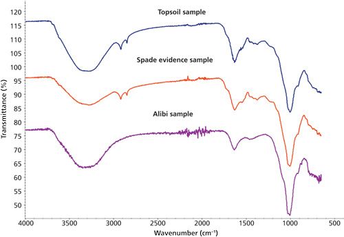
The top soil sample at the crime scene was highly organic, with the subsoil sample having a much lower proportion of organic matter present. The IR spectrum of the soil from the suspect’s spade also showed a relatively high proportion of SOM in the IR spectrum, although the intensity was slightly less than for the top soil sample at the crime scene. The pattern of bands from SOM in both the topsoil and spade IR spectra was very similar. The alibi sample had limited SOM compared with the crime scene samples and its chemical profile was different, indicating more humified material present within it (see Figure 6). The variable strength of the water peaks in the spectra did not interfere with the pattern recognition. The spectra from the arable and improved grassland sites showed that they were low in SOM. The only comparison site where the IR spectra showed appreciable SOM was the unimproved grassland, and in those samples the SOM pattern appeared different from that of the crime scene and the suspect’s spade.
Conclusions
It was possible for good IR spectra to be achieved in situ, with clear detail of the SOM content and the clay minerals, although further development of the method is necessary to produce consistently good spectra.
When the IR spectrum of the sample from the questioned spade was compared to the IR spectra of the other samples collected it was shown to be very similar to that of the top soil at the crime scene, with appreciable differences to the alibi, so there was a strong likelihood that the spade had been used at the crime scene.
Based on the results of this preliminary study, it appears that handheld FT-IR has considerable potential for in situ analysis of soil for forensic applications, even in a situation where the underlying geology is essentially very similar.
Acknowledgments
We would like to thank Tony Fraser for his expert help in reviewing this article. We would also like to acknowledge funding from the Scottish Government RESAS Department and from the EU MiSAFE project for the work described in the article.
Please note that 2015 is the International Year of Soils. For more information please visit www.fao.org/soils-2015/en/.
References
(1) L.A. Dawson and S. Hillier, Surf. Interface Anal.42, 363-377 (2010).
(2) R.W. Fitzpatrick, M.D. Raven, and S. Forrester in Criminal and Environmental Soil Forensics, K. Ritz, L. Dawson, and D. Miller, Eds. (Springer Netherlands, 2009), pp. 105-12.
(3) N.C. Brady and R.R. Weil, The Nature and Properties of Soils (Prentice Hall, New Jersey, 1996).
(4) R.E. Grim, Clay Mineralogy (McGraw Hill Book Company, Inc., New York, 2nd Edition, 1968), pp. 503-528.
(5) A.C.D. Newman, Chemistry of Clays and Clay Minerals, A.C.D. Newman, Ed. (Longman Scientific and Technical, Mineralogical Society, 1987), pp. 237-274.
(6) W.B. White in The Infrared Spectra of Minerals, V.C. Farmer, Ed. (Mineralogical Society, London, 1974), pp. 227-284.
(7) J.D. Russell and A.R. Fraser in Clay Mineralogy: Spectroscopic and Chemical Determinative Methods, M.J. Wilson, Ed. (Chapman and Hall, London, 1994), pp. 11-67.
(8) R.R.E. Artz, S.J. Chapman, J. Robertson, J.M. Potts, F. Laggoun-Defarge, S. Gogo, L. Comont, J.-R. Disnar, and A.-J. Francez, Soil Biol. Biochem.40, 515-527 (2008).
(9) http://www.macaulay.ac.uk/soilfit.
(10) A.H.J. Robertson, S.J. Hillier, C. Donald, H.R. Hill, and The NSIS Team, “A Robust FTIR Database for Scotland,” in Proceedings of the 3rd Global Workshop on Proximal Soil Sensing, May 26-29, 2013, Potsdam, Germany, pp. 182-185 (2013).
(11) B. Stenberg, R.A. Viscarra Rossel, A.M. Mouazen, and J. Wetterlind, in Advances in Agronomy, D.L. Sparks, Ed. (Elsevier Academic Press Inc., San Diego, California, 2010), pp. 163-215.
(12) T. Nguyen, L.J. Janik, and M. Raupach, Aust. J. Soil Res.29(1), 49-67 (1991).
(13) A.H.J. Robertson, A.M. Main, L.J. Robinson, and L.A. Dawson, “In Situ FTIR Analysis of Soil Samples at a Mock Crime Scene,” in Proceedings of the 4th Global Workshop on Proximal Soil Sensing, May 12-15, 2015, Hangzhou, China, pp. 12-17 (2015).
(14) http://forensicmisafe.wix.com/misafe.
A.H. Jean Robertson, Angela M. Main, Lucinda J. Robinson, and Lorna A. Dawson are with The James Hutton Institute in Craigiebuckler, Aberdeen, Scotland. Direct correspondence to: jean.robertson@hutton.ac.uk

AI Shakes Up Spectroscopy as New Tools Reveal the Secret Life of Molecules
April 14th 2025A leading-edge review led by researchers at Oak Ridge National Laboratory and MIT explores how artificial intelligence is revolutionizing the study of molecular vibrations and phonon dynamics. From infrared and Raman spectroscopy to neutron and X-ray scattering, AI is transforming how scientists interpret vibrational spectra and predict material behaviors.
Real-Time Battery Health Tracking Using Fiber-Optic Sensors
April 9th 2025A new study by researchers from Palo Alto Research Center (PARC, a Xerox Company) and LG Chem Power presents a novel method for real-time battery monitoring using embedded fiber-optic sensors. This approach enhances state-of-charge (SOC) and state-of-health (SOH) estimations, potentially improving the efficiency and lifespan of lithium-ion batteries in electric vehicles (xEVs).
New Study Provides Insights into Chiral Smectic Phases
March 31st 2025Researchers from the Institute of Nuclear Physics Polish Academy of Sciences have unveiled new insights into the molecular arrangement of the 7HH6 compound’s smectic phases using X-ray diffraction (XRD) and infrared (IR) spectroscopy.