Synthesis of Differently Sized Gold Nanoparticles for SERS Applications in the Detection of Malachite Green
Malachite green (MG) is a water pollutant that is difficult to detect rapidly. In this study, surface-enhanced Raman spectroscopy (SERS) was used to detect the presence of MG in water. Three kinds of gold nanoparticles with different sizes were prepared via a traditional sodium citrate reduction method and used for the SERS detection of MG standard samples. Optimally sized gold nanoparticles were used as the SERS substrate for the detection of MG in simulated real water samples. The SERS enhancement effect was the greatest when using gold nanoparticles with sizes of 36±4 nm. In the simulated real water samples, the minimum detection limit of MG was 10-7 M.
Fish in a culture fishery have a higher risk of infection by various bacteria, fungi, and parasites. Water mildew, cucurbitacin, and various high-risk diseases threaten the growth of fish (1–3). Fishermen add malachite green (MG) to the body of water to mitigate the effects of bacteria and parasites on freshwater aquatic products, keeping fish healthy. MG is a synthetic triphenylmethane chemical that serves as an antibiotic; however, its use has been prohibited due to its toxicity (4). Also, MG induces side effects, such as deformity and gene mutation. When it enters the body, MG is metabolized into toxic substances that are extremely harmful to human health (5). The amount of MG ingested at one time may not exceed the standard, and there may be no distinct symptoms of poisoning. However, MG remains in the human body for a long time, and, after accumulating to a certain concentration, it may cause various diseases (6,7). Furthermore, the long-term accumulation of MG could lead to cancer; therefore, the detection of MG is of great significance. There are many methods to detect MG, including high performance liquid chromatography (HPLC), thin-layer chromatography, liquid chromatography–mass spectrometry (LC-MS), and enzyme-linked immunoassay (8–12). These methods are not suitable for rapid detection, as they involve many experimental steps, complex operation, expensive equipment, and require highly skilled operators.
Raman spectroscopy is a commonly used analytical method in molecular structure research and has advantages and disadvantages. The Raman scattering signal representing the structural characteristics of the measured material is relatively weak, and is difficult to detect. Additionally, the Raman fingerprint peak is scattered when the concentration of the tested sample is low, making it difficult to detect the Raman signal, affecting the accuracy and feasibility of the experiment (13–15). Reliability limits the application of Raman spectroscopy in high-sensitivity detection and analysis. Surface-enhanced Raman spectroscopy (SERS) is a special Raman scattering phenomenon in which sample molecules are adsorbed on the surface of certain materials (such as Au and Ag) to enhance the Raman scattering intensity of molecules (3,16,17). SERS has been widely used as a material analysis and testing technology, given its non-destructiveness at the molecular level, high sensitivity, and good resolution, without requiring special sample treatment (5,18,19).
Noble metal nanoparticles are used in many fields, such as photosensitivity, catalysis, photoelectronics, information storage, and SERS, due to their unique characteristics and excellent properties (13,20,21). Among them, gold nanoparticles have stable properties, require a simple preparation process, and can be synthesized with a uniform particle size. Gold nanoparticles can not only be used as a research object in many experiments, but also as a SERS substrate. Different preparation methods and related properties of gold nanoparticles are outlined in the literature (22,23).
In this study, gold nanoparticles with different particle sizes were prepared by a sodium citrate reduction. They were characterized by ultraviolet–visible spectroscopy (UV-vis) and scanning electron microscopy (SEM) to determine the maximum absorption spectrum wavelength and particle size range. Crystal violet (CV) was used as a probe to detect the sensitivity and reproducibility of the three substrates. The three substrates were then used to detect different concentrations of MG via SERS, in both standard samples and actual water samples.
Materials and Methods
Chemical Materials
HAuCl4·3H2O, sodium citrate, ʟ-ascorbic acid, NaBH4, AgNO3, H2O2, and MG were purchased from China National Medicine Chemical Reagent Co., Ltd. All chemicals were of analytical grade, and their solutions in distilled water were prepared without further purification. Actual water samples were taken from the pond at the Hefei Institute of Physical Sciences at the Chinese Academy of Sciences. Double-distilled water (>18.0 MΩ cm) obtained using a Millipore Milli-Q Gradient system was used for all experiments.
Characterization
The morphology and structure of the nanoparticles were observed by field-emission scanning electron microscopy (FESEM; Sirion-200, FEI) using a field-emission gun operating at 200 kV. Raman spectra were recorded on a LabRam HR-800 Raman spectrometer (Horiba Jobin Yvon). An excitation wavelength of 633 nm and an integration time of 3 s was used for all Raman tests. UV-vis absorption spectra were recorded on a UV-2550 spectrophotometer (Shimadzu).
Synthesis of Au Nanoparticles
Gold nanoparticles with different sizes were prepared by a sodium citrate reduction. For this, 1 mL of 1% AuCl4·3H2O and 99 mL of ultrapure water were added to three clean flasks, then stirred and heated on a temperature-controlled magnetic stirrer (heated by a heating jacket and air bath). When the solution began to boil, 1 mL, 2 mL, or 5 mL of sodium citrate was rapidly added to the three flasks while stirring. After 30 min of heating, the gold nanoparticles were cooled until they reached approximately 27–29 °C. Three kinds of wine-red gold nanoparticles with a HAuCl4·3H2O to sodium citrate volume ratio of 1:1, 1:2, and 1:5 were obtained, referred to as E1, E2, and E3, respectively.
Sample Preparation and SERS Measurements
Sample Preparation
Three different gold-nanoparticle solutions of different ratios were prepared. A 1 mL aliquot of each solution was subjected to centrifugation at 6000 rpm for 10 min, after which the excess surfactant and supernatant were removed. Warm ultrapure water was subsequently added to the centrifuge tubes, followed by centrifugation under the same conditions for 10 min. After removal of the supernatant, the gold nanoparticles that were dispersed in water after centrifugation were self-assembled onto a clean silicon wafer for use.
SERS Measurements
All samples were analyzed using a LabRAM HR800 confocal microscope Raman system (Horiba Jobin Yvon) with a 633 nm He-Ne laser source. The Raman spectra were collected between 200 and 2000 cm-1, and the integration time for each spectrum was 3 s.
Results and Discussion
Optical Characterization of Different-sized Gold Nanoparticles
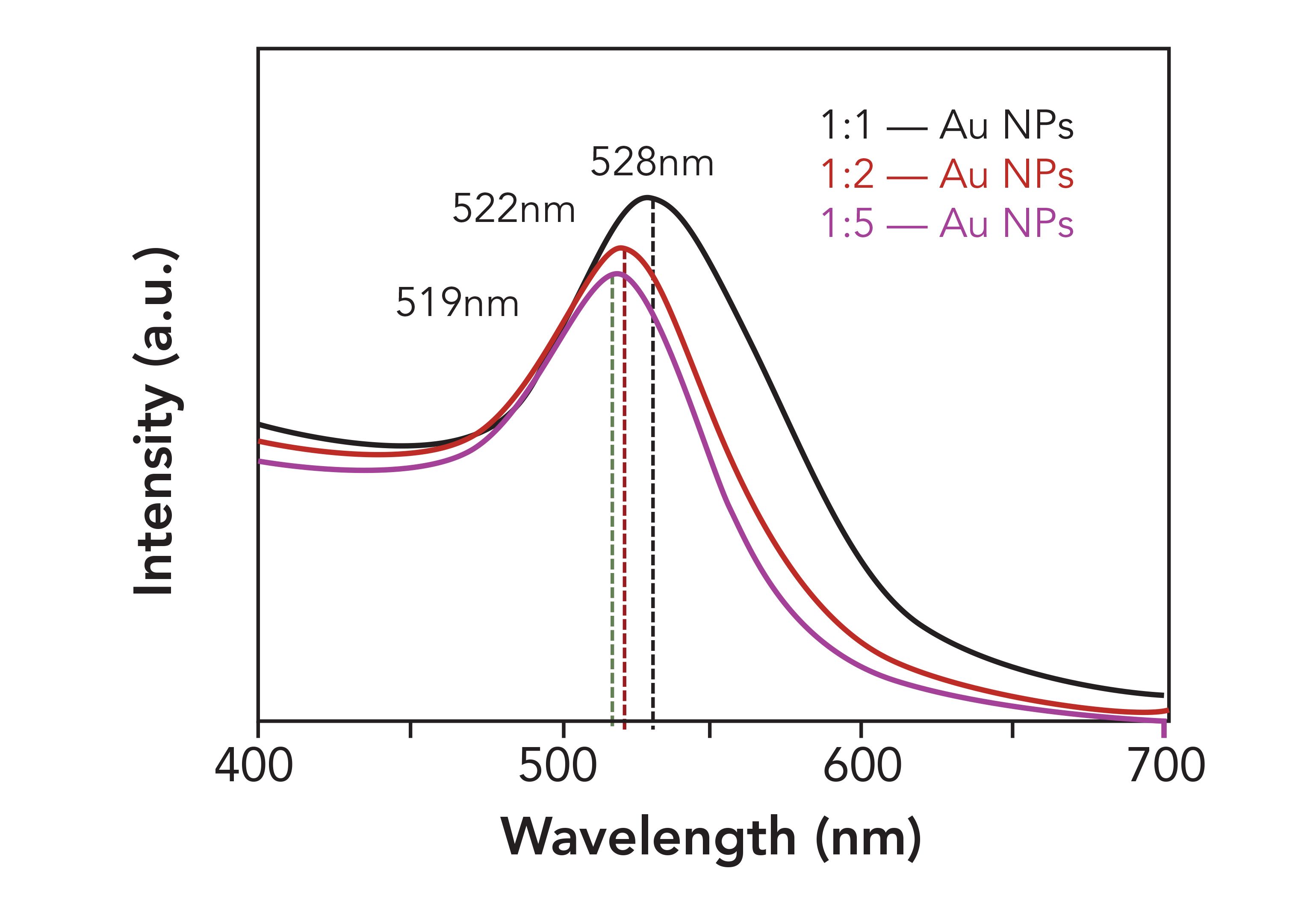
Surface plasmon absorption peaks typically occur for gold nanoparticles at 520 nm, and the positions of these peaks vary with the size and morphology of the gold nanoparticles. Therefore, UV-vis absorption spectra of gold nanoparticles can reflect the size characteristics of their morphologies and particles (24). Figure 1 shows the UV-vis absorption spectra of three sizes of gold nanoparticles. The maximum absorption peaks of E1, E2, and E3 occur at 519, 522, and 528 nm, respectively. The experimental results show that the maximum absorption peaks are red-shifted with the decreasing ratio of sodium citrate in the system.
FIGURE 1: UV-vis spectra of gold nanoparticles with different sizes.
To determine the size and morphology of the three gold nanoparticles, SERS substrates with these nanoparticles were analyzed by SEM. Figures 2a, 2b, and 2c show that the sizes of E1, E2, and E3 range from 36±4, 18±4, and 15±2 nm, respectively. Hence, E1 has the largest size. The images show that gold nanoparticles with the adsorbed ordered monolayer were formed without distinct agglomeration. It can be observed that the gold nanoparticles are closely arranged and uniform in shape, and therefore, can be used as a substrate for SERS detection.
FIGURE 2: SEM images of gold nanoparticles with different particle sizes; (a) E1, (b) E2, (c) E3.

Sensitivity of Gold Nanoparticle SERS Substrates
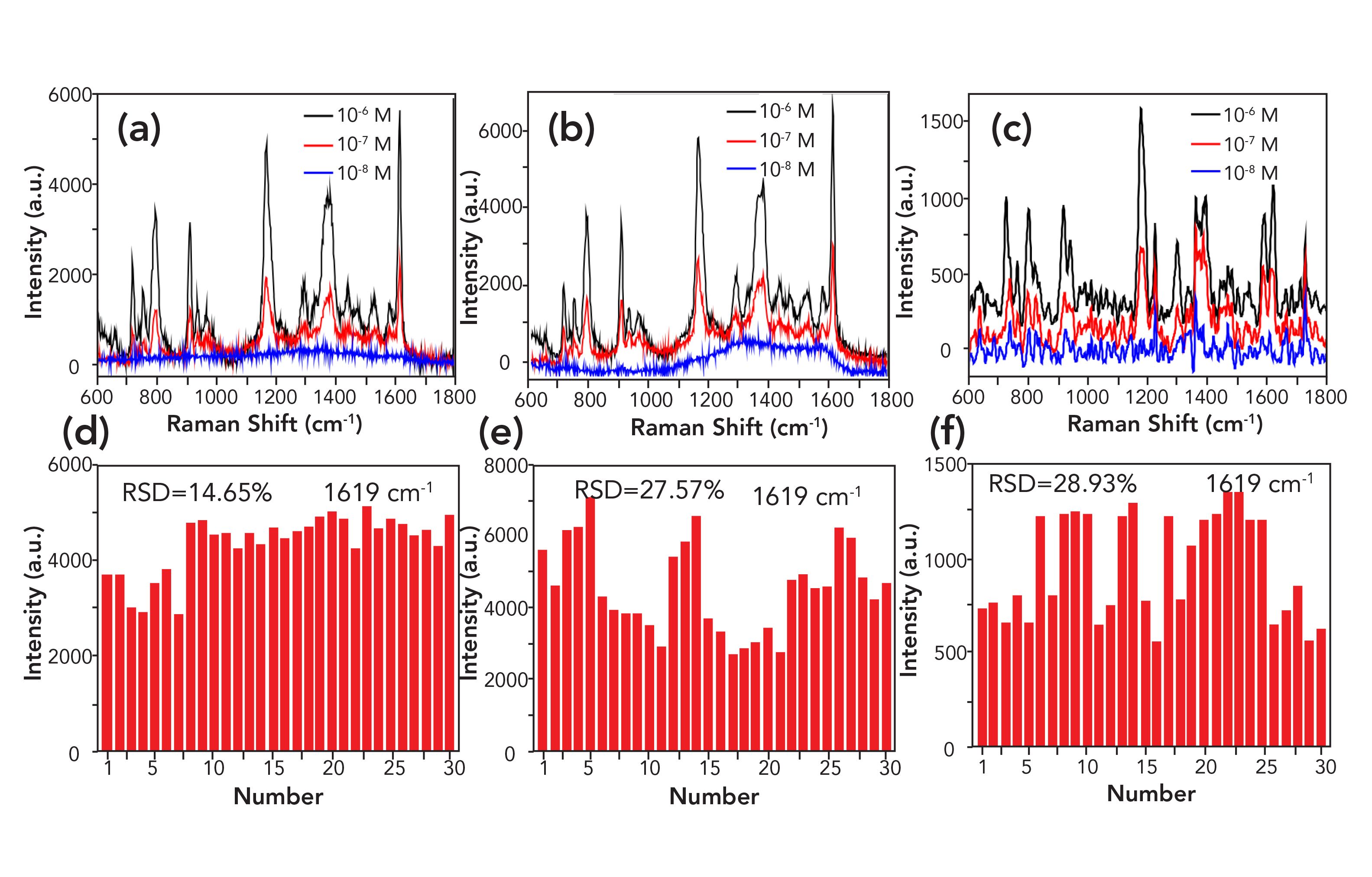
To evaluate their sensitivity as SERS substrates, the self-assembled, differently-sized gold nanoparticles were tested using the CV solution as a probe (25). The three kinds of gold nanoparticles were centrifuged. Standard samples of the CV solution with concentrations of 10-6 M, 10-7 M, or 10-8 M were evenly mixed with 2 μL of the standard sample solution each in a 1:1 ratio, and then dropped on a clean silicon wafer. After air drying, the samples were tested using a Raman spectrometer. Figure 3 shows that the Raman fingerprint peaks of the CV molecules occur at 1619 cm-1, 1370 cm-1, and 1171 cm-1. The gold nanoparticle SERS for the E1 substrate exhibits a strong detection signal for CV, with concentrations of 10-6 M and 10-7 M. No signal was detected at 10-8 M of the CV solution, indicating that the detection limit for CV was 10-7 M. As seen in Figures 3b and 3c, the SERS for the E2 and E3 substrates were observed to generate a strong detection signal for CV, with concentrations of 10-6 M and 10-7 M. This signal was not distinct for 10-8 M, indicating that the detection limit for the standard CV samples is 10-7 M.
Figures 3d, 3e, and 3f are the relative standard deviations (RSD) of the 30 points that were calculated for the 10−6 M CV solution, corresponding to the 1619 cm−1 fingerprint peaks. The RSD for the E1 substrate was 14.65% (<20%). The results showed that the E1 substrate has good reproducibility. As shown in Figures 3e and 3f, the RSDs for E2 and E3 are 27.57% and 28.93%, respectively (>20%), which indicate that the E2 and E3 substrates are not evenly distributed, and do not have a suitable signal enhancement effect on the target molecules. Therefore, these may not be suitable for use as substrates.
FIGURE 3: SERS spectra of CV, at different concentrations, collected on three gold nanoparticles with (a) E1, (b) E2, and (c) E3. (d–f) Respective RSDs of the main CV (10−6 M) vibration at 1619 cm−1 intensities determined for 30 spot locations.
SERS Detection for MG Standard Sample
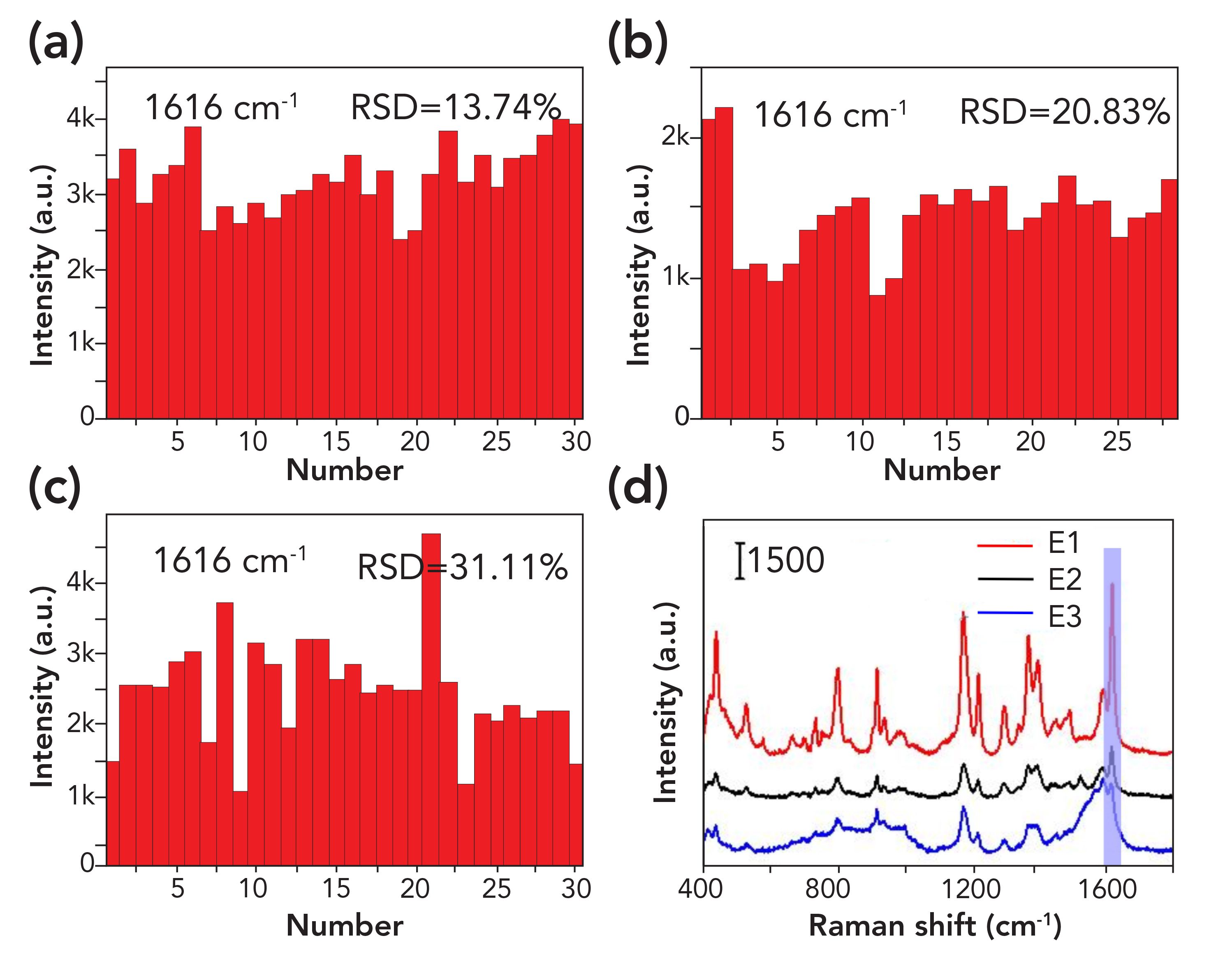
By comparing the SERS spectra of 10-5 M MG standard samples with E1, E2, and E3 as substrates in Figure 4d, it was found that E1 led to the greatest SERS enhancement effect. From Figures 4a, 4b, and 4c), the RSDs of the SERS intensity of the three substrates at 1616 cm-1 were calculated to be 13.74%, 20.83%, and 31.11%, respectively. Therefore, the E1 substrate has good reproducibility and high stability. The reproducibility of E2 was acceptable and E3 was poor and is unstable. Hence, the detection effect of E1 for MG was the best.
FIGURE 4: (a–c) SERS intensities for the three sizes (E1, E2, E3, respectively) of gold nanoparticles at 1616 cm-1 and (d) SERS spectra of 10-5 M for MG samples obtained using the three substrate samples.
To further evaluate the detection effect of the three substrates on MG, we conducted sensitivity and reproducibility experiments of MG, as shown in Figures S1, S2, and S3. Figures S1a, S2a, and S3a show that the intensity of the SERS spectra decreased with decreasing MG concentration (10-5 to 10-7 M), and the MG detection limit was identified as 10-7 M. The above spectra featured several MG-derived peaks (908, 1170, 1295, 1394, and 1616 cm-1), the positions of which were shifted slightly compared with values reported in literature (23). Under the same detection conditions, the detection signal strength of E3 was the lowest.
FIGURE S1: Detection of MG standard solution using E1 gold nanoparticles as a substrate. (a) SERS spectra of different MG concentrations. (b) SERS spectra of 10−5 M for MG from five independent SERS measurements. (c) and (d) show the intensities of MG solution (at 10−5 M) in the 30 spots SERS line-scan spectra collected on the substrate at 1170 cm-1, and 1394 cm-1, respectively.
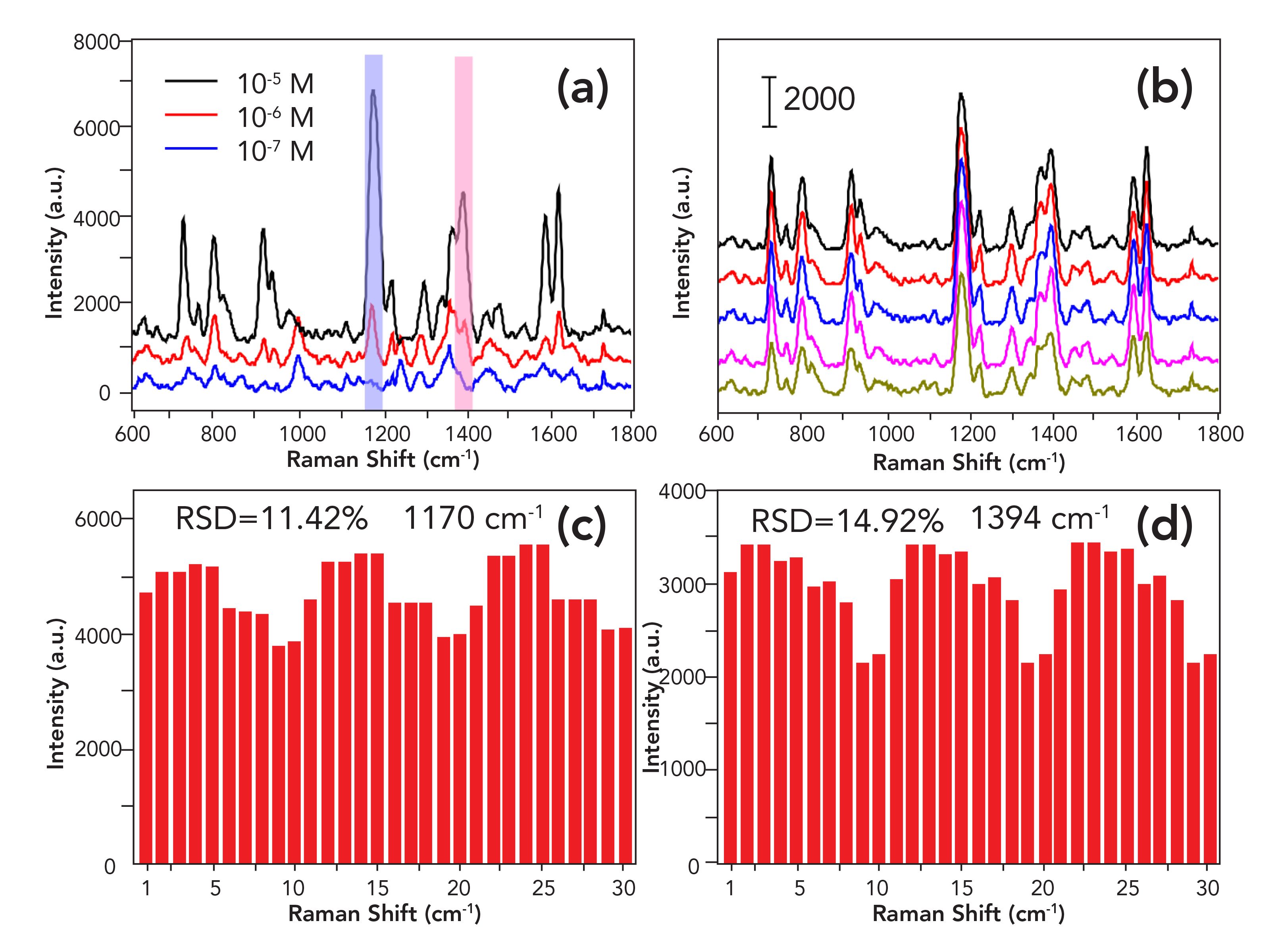
FIGURE S2: Detection of MG standard solution using E2 gold nanoparticles as a substrate. (a) SERS spectra of different MG concentrations. (b) SERS spectra of 10−5 M for MG from five independent SERS measurements. (c) and (d) show the intensities of MG solution (at 10−5 M) in the 30 spots SERS line-scan spectra collected on the substrate at 1170 cm-1, and 1394 cm-1, respectively.
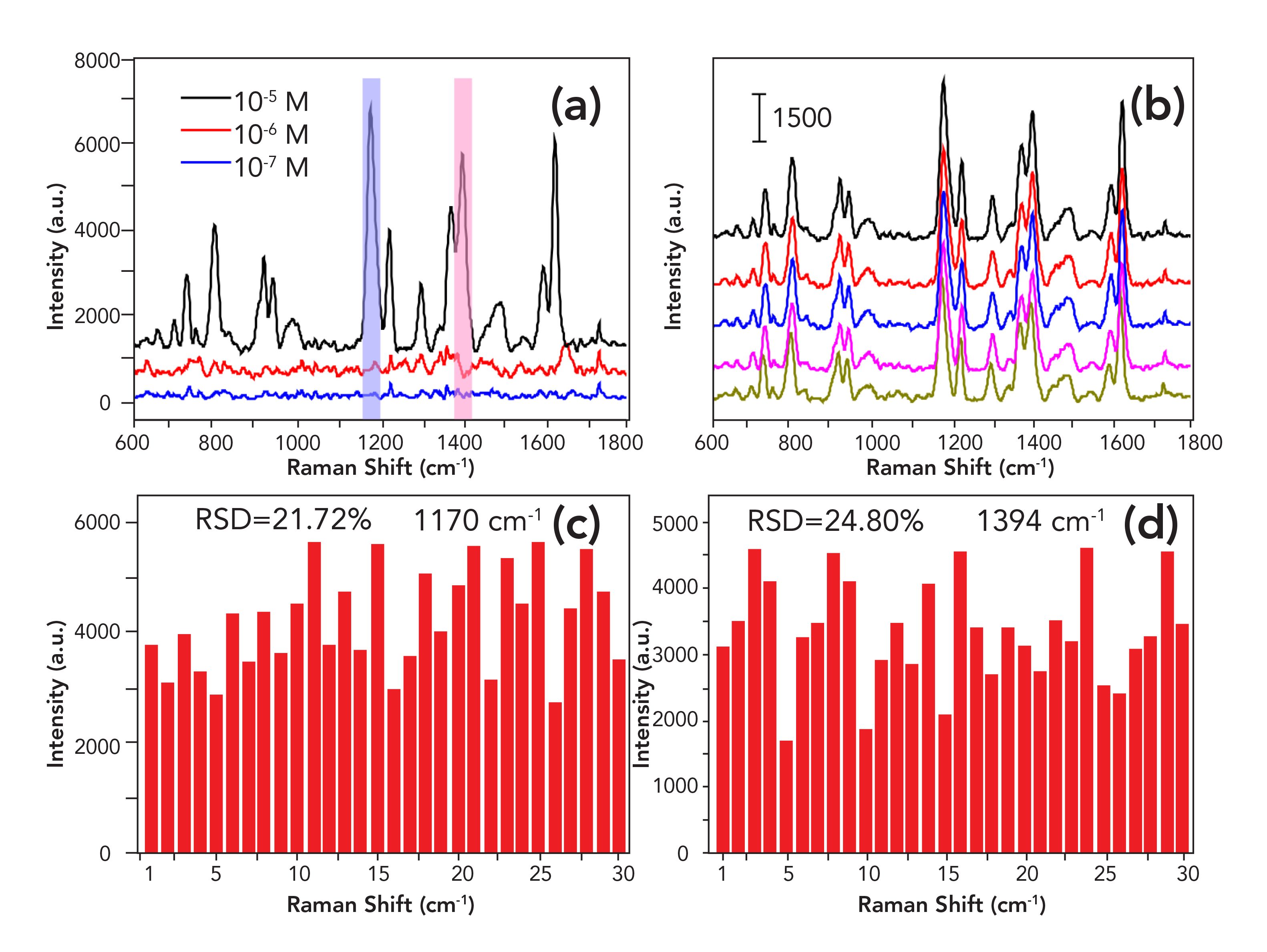
FIGURE S3: Detection of MG standard solution using E3 gold nanoparticles as a substrate.(a) SERS spectra of different MG concentrations. (b) SERS spectra of 10−5 M for MG from five independent SERS measurements. (c) and (d) show the intensities of MG solution (at 10−5 M) in the 30 spots SERS line-scan spectra collected on the substrate at 1170 cm-1, and 1394 cm-1, respectively.
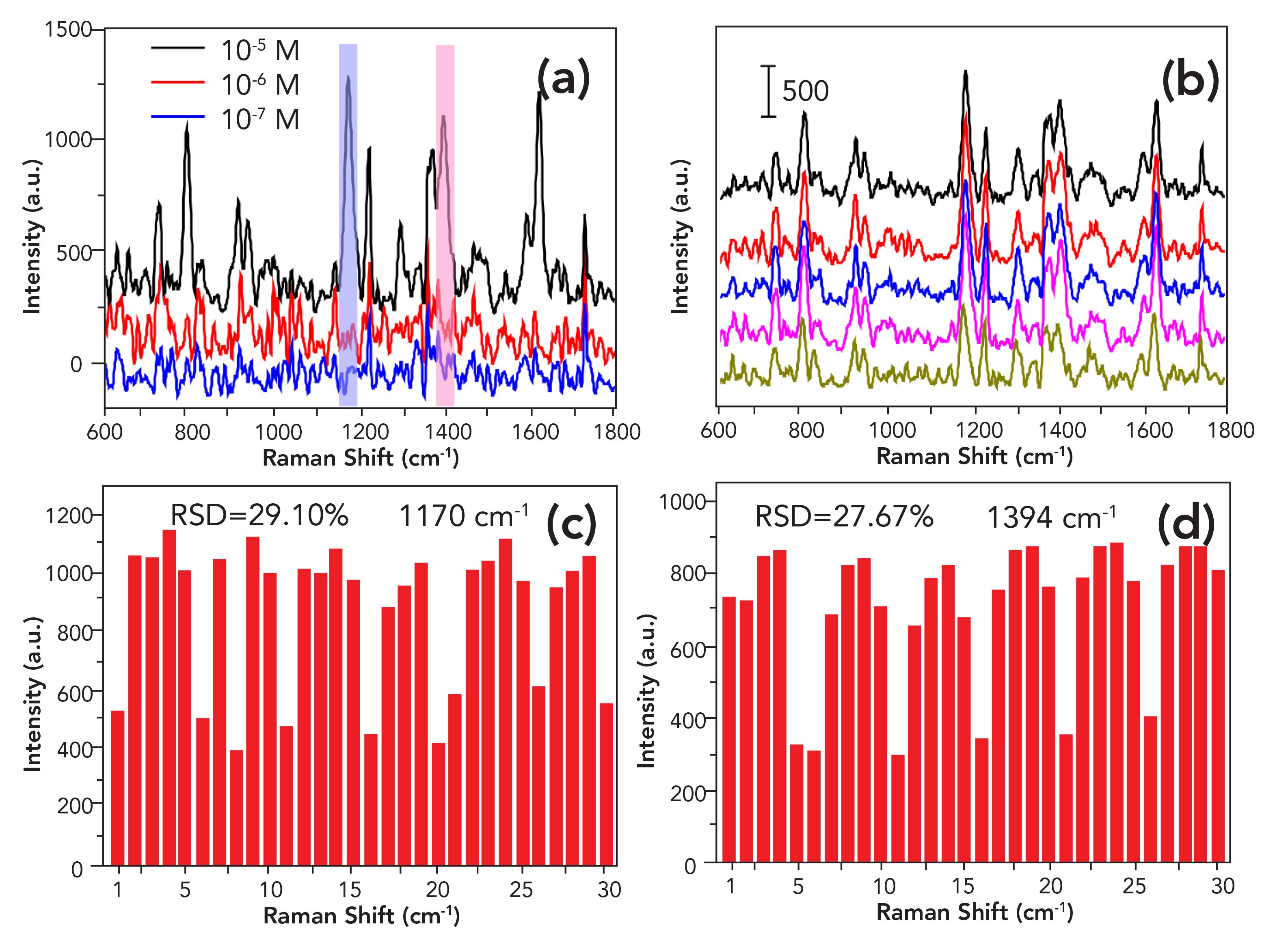
Figures S1, S2, and S3 show the RSDs of the 30 points that were calculated for the 10−5 M MG solution, corresponding to the 1170 cm−1 and 1394 cm-1 fingerprint peaks. The RSDs in S1 are 11.42% and 14.99% (less than 20%). The results showed that the E1 substrate has good reproducibility. As shown in Figures S2 and S3, the RSDs for E2 and E3 are 21.72% for 1170 cm-1 (24.80% for 1394 cm-1) and 29.10% for 1170 cm-1 (27.67% for 1394 cm-1), respectively. Consequently, this indicates that the E2 and E3 substrates are not evenly distributed, and do not exhibit a good signal enhancement effect on the target molecules; therefore, these may not be suitable for use as substrates.
From the above discussion, the E1 substrate has the best sensitivity and reproducibility, and the SERS signal of the MG standard sample is the largest. Therefore, we chose E1 as the substrate for the SERS of MG simulated real water samples.
SERS Detection of MG Simulated Real Water Samples
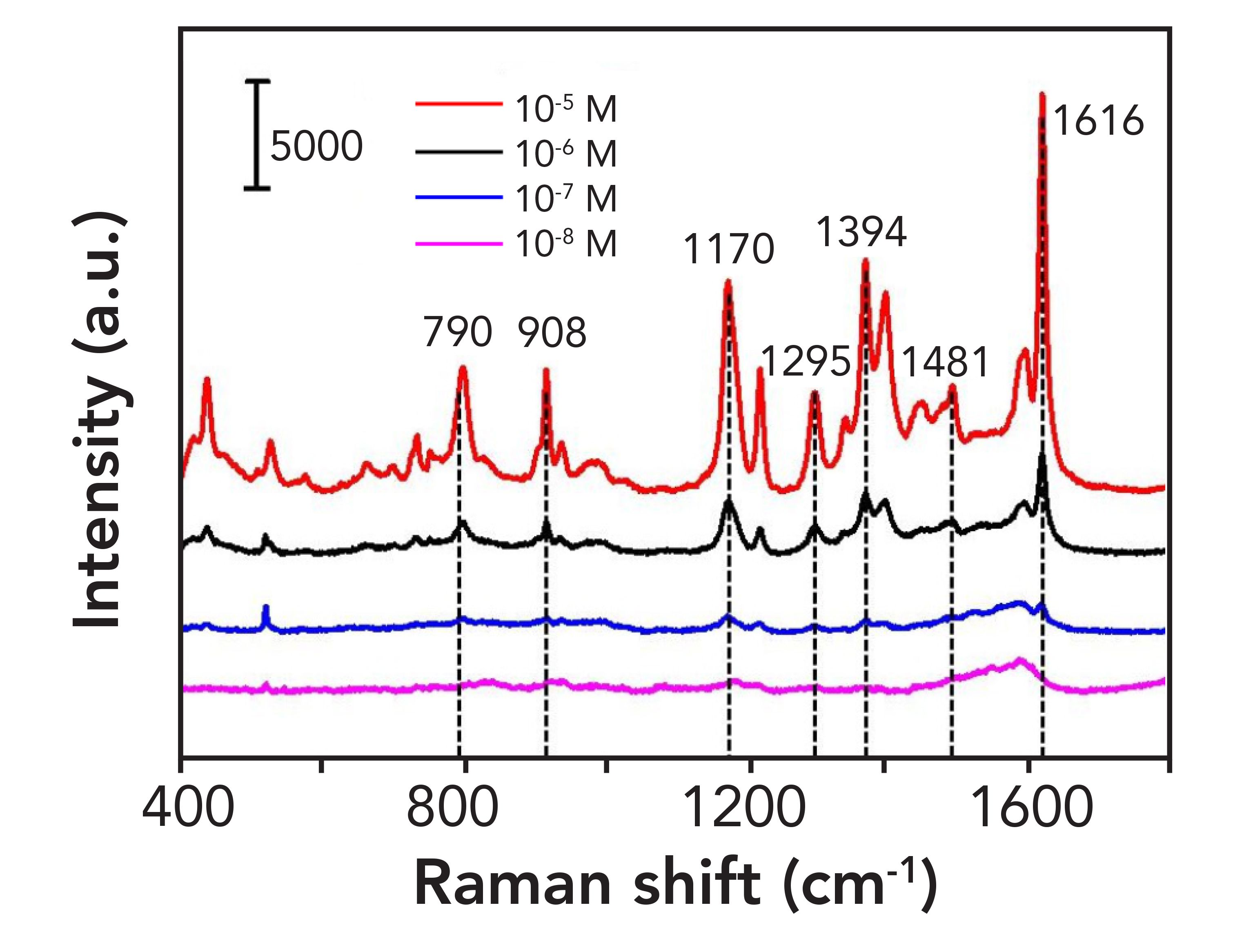
E1 gold nanoparticles were mixed with simulated water samples at concentrations of 10-5 M, 10-6 M, 10-7 M, and 10-8 M MG at a ratio of 1:1, dripped on to clean silicon wafers, and dried. Later, they were analyzed by a Raman spectrometer. The bands at 1616, 1481, and 1295 cm-1 are assigned to the out-of-plane vibration of aromatic rings. The peaks at 1394 cm-1 correspond to the bending vibration in the C-H plane, and the strong peaks at 1170 cm-1 represent bending vibrations in the C-H plane of aromatic rings. The bands at 908, 1170, and 1394 cm-1 are fingerprint peaks of MG (4); the assignments of the major Raman peaks of MG are summarized in Table I, although it should be noted that the positions of the peaks were slightly shifted compared with those reported in the literature (23,26). Figure 5 shows that the intensity of the SERS spectra decreased with diminishing MG concentration (10-5 to 10-8 M), with no band and fingerprint peaks observed at 10-8 M. This indicates that the detection limit of MG in real water samples can reach 10-7 M on E1 substrates.
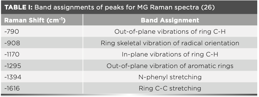
FIGURE 5: SERS spectra for different concentrations of MG in simulated real water samples.
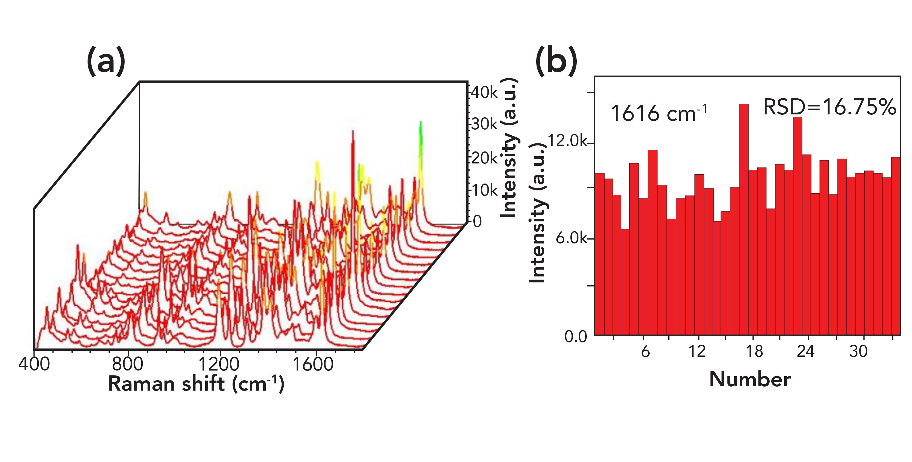
In the SERS analysis of the 10-5 M MG simulated water sample using E1 as the substrate, the SERS signal intensity was high, and the enhancement effect of SERS was distinct. Figure 6b shows that the intensity of the Raman fingerprint peak is similar for the same Raman wave number. The RSD of the SERS intensity at several points of the 1616 cm-1 fingerprint peak was calculated to be 16.75% (<20%), indicating that the substrate has good reproducibility. Combining the results from Figures 6a and 6b, it can be concluded that the 1:1 gold-nanoparticle substrate has good reproducibility. The SERS substrate has good orderliness, and a uniform surface enhancement effect. The SERS intensity of MG is high, with a distinct enhancement effect, and the Raman spectra are stable. The SERS spectra of the 10-5 M MG standard samples and simulated real water samples show that the SERS intensity of the latter is weak, because of other interfering factors affecting the Raman signal of MG in real water.
FIGURE 6: (a) 3D SERS spectrogram of 10-5 M for MG simulated water sample; (b) SERS intensity from SERS spectrum at 1616 cm-1.
Conclusions
Three kinds of gold nanoparticles with different diameters were synthesized by a sodium citrate reduction. The nanoparticles were employed for SERS detection of MG in a standard sample, and it was concluded that the sample with 1:1 gold nanoparticles had the best detection effect. These nanoparticles were employed for the SERS detection of MG in simulated real water samples, where the detection limit reached 10-7 M. SERS detection is advantageous, owing to its high recognition ability, high sensitivity, good reproducibility, and simple operation. The SERS detection of MG is a simple method that exhibits a remarkable sensitivity for standard solution detection, an enhanced practical application value, and can achieve a high-sensitivity detection of MG in a real water sample.
Funding
This work was supported by the Natural Science Research Key Project of Education Department of Anhui Province (No. KJ2019A0669 and No. KJ2019A0671), Daze Scholar Project (No. 2018szxydzxz02), Key Discipline of Material Science and Engineering of Suzhou University (No. 2017XJZDXK3), and Natural Science Research Key Project of Education Department of Anhui Province (No. KJ2018A0454).
References
(1) C.Y. Huang and C.H. Chien, Appl. Sci.-Basel. 23, 9–10 (2019).
(2) R. Wang, L.P. Zhang, S.Y. Zou, and H. Zhang, Microchem. 150, 104127–104131 (2019).
(3) M.L. Mekonnen, W.N. Su, C.H. Chen, and BJ. Hwang, Anal. Meth. UK 48, 6823–6829 (2017).
(4) L. Ouyang, L. Yao, T.H. Zhou, and L.H. Zhu, Anal. Chim. Acta 1027, 83–91 (2018).
(5) T.T. Xu, X.H. Wang, Y.Q. Huang, K.Q. Lai, and Y.X. Fan, Food Control 106, 106720 (2019).
(6) S.X. Guo, F.L. Zhang, P. Gao, Y.M. Zeng, H.J. Chen, G.K. Liu, and L. Wang, Spectrosc. Spect. Anal. 34(5), 1284–1288 (2014).
(7) N.N. Xu, Q. Zhang, W. Guo, Q.T. Li, and J. Xu, Chin. J Anal. Chem. 44(9), 1378–1383 (2016).
(8) A.A. Bergwerff and P. Scherpenisse, J. Chromatogr. B 788(2), 351–359 (2003).
(9) K. Mitrowska, A. Posyniak, and J. Zmudzki, J. Chromatogr A 1089(1–2), 187–192 (2005).
(10) R.J. Teichman, G.H. Takei, and J.M. Cummins, J. Chromatogr. 88(2), 425–427 (1974).
(11) M.R. Khan, S.M. Wabaidur, R. Busquets, M.A. Khan, M.R. Siddiqui, and M. Azam, Process Saf. Environ. 126, 160–166 (2019).
(12) Y. Wang, J.Y. Yang, Y.D. Shen, Y.M. Sun, Z L. Xiao, H.T. Lei, H. Wang, and Z.L. Xu, Food Agr. Immunol. 28(6), 1460–1476 (2017).
(13) G.D. Fu, D.W. Sun, H.B. Pu, and Q.Y. Wei, Talanta 195, 841–849 (2019).
(14) H.P. Fu, J.M. Chen, L.J. Chen, X. Zhu, Z.L. Chen, B. Qiu, Z.Y. Lin, L.H. Guo, and G.N. Chen, Microchim. Acta 186(2), 1–7 (2019).
(15) X.M. Nie, J. Wang, X. Wang, Y.P. Tian, S. Chen, Z.Y. Long and C.H. Zong, Chin. J. Chem. Phys. 32(4), 444–450 (2019).
(16) L. Sun, M. Zhang, V. Natarajan, X.F. Yu, X.L. Zhang, and J.H. Zhan, RSC Adv. 7(38), 23866–23874 (2017).
(17) N.H. Ly, C.H. Seo, and S.W. Joo, Sensors-Basel 16(11), 1785–1795 (2016).
(18) A. Kumar and V. Santhanam, Anal. Chim. Acta 1090, 106–113 (2019).
(19) G.N. Xiao, L. Li, A.M. Yan, and X.Y. He, Spectrochim. Acta A 223, 117269–117274 (2019).
(20) N. Yang, T.T. You, Y.K. Gao, C.M. Zhang, and P.G. Yin, J. Agr. Food Chem. 66(26), 6889–6896 (2018).
(21) Y.D. Lu, C.J. Wu, Y. Wu, R.Y. You, G. Lin, Y.Q. Chen and S.Y. Feng, Materials 11(7), 1197–1207 (2018).
(22) W. Li, X.G. Fan, X. Wang, M. Tang, J. Que, J. He, and Y. Zuo, Spectrosc. Spect. Anal. 37(6), 1778–1783 (2017).
(23) N.N. Xu, Q. Zhang, W. Guo, Q.T. Li, and J. Xu, Chin. J. Anal. Chem. 44(9), 1378–1383 (2016).
(24) Y. Jin, P.Y. Ma, F.H. Liang, D.J. Gao, and X.H. Wang, Anal. Methods-U. 5(20), 5609–5614 (2013).
(25) X.W. Chen, T.H.D. Nguyen, L.Q. Gu, and M.S. Lin, J. Food Sci. 82(7), 1640–1646 (2017).
(26) L.L. He, N.J. Kim,H. Li, Z.Q. Hu, and M.S. Lin, J. Agric. Food Chem. 56(21), 9843–9847 (2008).
Miao Qin, Cong Wang, Jun Zhu, Li Yong, and Hongyan Wang are with the Key Laboratory of Spin Electron and Nanomaterials of Anhui Higher Education Institutes, at the School of Chemistry and Chemical Engineering, at Suzhou University, in Suzhou, People’s Republic of China. Liangbao Yang is with the Center of Medical Physics and Technology at the Hefei Institutes of Physical Science, at the Chinese Academy of Sciences, in Hefei, People’s Republic of China. Direct correspondence to: lbyang@iim.ac.cn

AI-Powered SERS Spectroscopy Breakthrough Boosts Safety of Medicinal Food Products
April 16th 2025A new deep learning-enhanced spectroscopic platform—SERSome—developed by researchers in China and Finland, identifies medicinal and edible homologs (MEHs) with 98% accuracy. This innovation could revolutionize safety and quality control in the growing MEH market.
New Raman Spectroscopy Method Enhances Real-Time Monitoring Across Fermentation Processes
April 15th 2025Researchers at Delft University of Technology have developed a novel method using single compound spectra to enhance the transferability and accuracy of Raman spectroscopy models for real-time fermentation monitoring.
Nanometer-Scale Studies Using Tip Enhanced Raman Spectroscopy
February 8th 2013Volker Deckert, the winner of the 2013 Charles Mann Award, is advancing the use of tip enhanced Raman spectroscopy (TERS) to push the lateral resolution of vibrational spectroscopy well below the Abbe limit, to achieve single-molecule sensitivity. Because the tip can be moved with sub-nanometer precision, structural information with unmatched spatial resolution can be achieved without the need of specific labels.