The 2020 Emerging Leader in Molecular Spectroscopy Award
Markita Landry, the winner of the 2020 Emerging Leader in Molecular Spectroscopy Award, works at the intersection of single-molecule biophysics and nanomaterial-polymer science to develop new tools for understanding biological systems.
This year’s molecular spectroscopy award recipient, Markita Landry, is an outstanding young researcher in spectroscopy, biotechnology, and nanotechnology. Landry works at the intersection of single-molecule biophysics and nanomaterial-polymer science and has developed new tools for understanding biological systems. Using a combination of nanoparticles, imaging, and spectroscopy, her work has led to the discovery of aspects of neuromodulation in the brain, and for the delivery of genetic materials into plants for crop biotechnology applications.
Markita Landry, PhD, is an assistant professor at University of California-Berkeley. She is the winner of the 2020 Emerging Leader in Molecular Spectroscopy Award, which is presented by Spectroscopy magazine. This annual award, selected by an independent scientific panel, recognizes the achievements and aspirations of a talented young molecular spectroscopist who has made strides in his or her career toward the advancement of molecular spectroscopy techniques or applications. The winner must be within 10 years of receiving his or her highest academic degree in the year the award is presented. The award was scheduled to be presented to Landry at the SciX 2020 conference in October, where she was to give a plenary lecture and be honored in an award symposium. Due to the COVID-19 pandemic, the conference is now scheduled to be a virtual one, and Landry’s award presentation will be part of that virtual event.
Landry received her PhD from the University of Illinois at Urbana-Champaign in 2012. She then served as a postdoctoral researcher at the Massachusetts Institute of Technology before taking her current position at the University of California Berkeley. She is also currently a faculty scientist with the Lawrence Berkeley National Laboratory, an investigator at the Chan-Zuckerberg Biohub in San Francisco, and an investigator with the Innovative Genomics Institute in Berkeley.
Landry’s research lies on the intersection of single-molecule biophysics and nanomaterial–polymer science, focusing on developing new tools to probe and characterize biological systems. Her research has generated nanoparticle–polymer conjugates for imaging and spectroscopic detection of neuromodulation in the brain and for the delivery of genetic materials into plants for crop biotechnology applications.
She has developed a near-infrared (NIR) fluorescent probe as a tool that enables imaging of the chemical communication between neurons in the brain at the spatial (micrometer) and temporal (millisecond) scales that the brain uses to communicate. This NIR nanoscale catecholamine probe molecule can be used to image the neuromodulator dopamine in the brain, and has been validated against “gold standard” techniques in neuroscience, including electrophysiology and optogenetics. Landry and her group also developed a generic, high-throughput approach for generating nanosensors for molecules for which molecular recognition elements do not exist. She and her team have demonstrated the utility of a single-walled carbon nanotube-based sensor platform to develop a serotonin nanosensor that can support explorations of serotoninergic imaging in the brain, as well as the effects of drugs at the level of individual nerve synapses, where signals pass from one neuron to another.
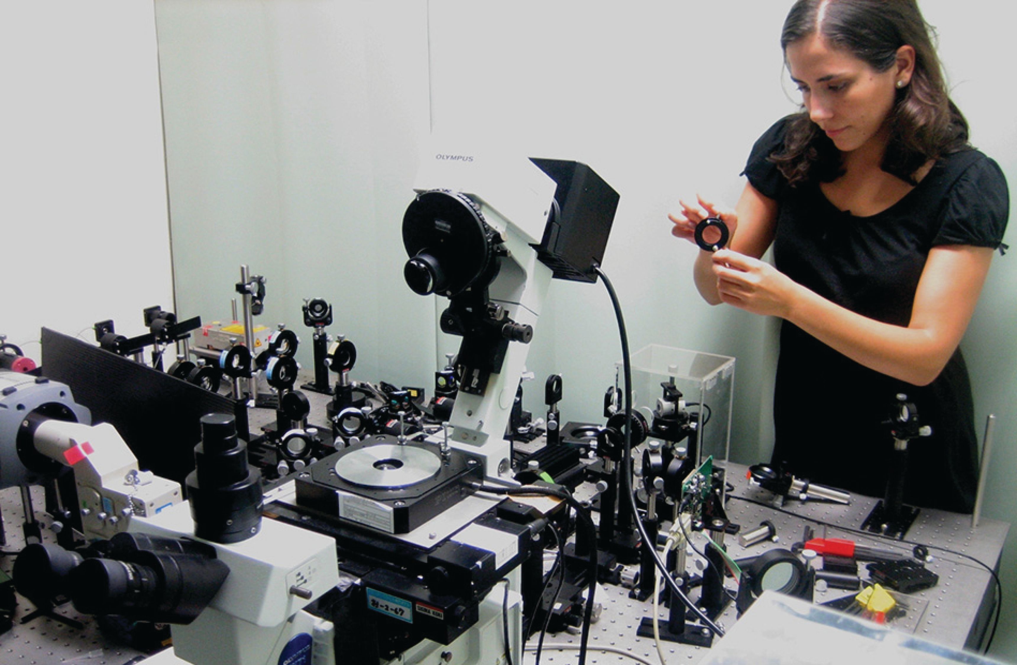
Figure 1: Landry in her laboratory with a single molecule microscope that her group uses for neuroimaging research designing experiments in single-walled carbon nanotube-based technologies. (Source: Photo courtesy of Prabir Patra)

In her plant research, Landry has developed a new approach to genetic engineering using nanomaterials and their associated surface chemistries to controllably graft and quickly deliver functional biomolecular cargoes, such as whole DNA plasmids to agriculturally relevant plants. These tools accomplish nanoparticle-mediated heterologous and transient expression of a protein in plants, and, unlike in genetically modified organisms (GMOs), there is no transgenic DNA integration into the host plant genome. This DNA plasmid delivery approach provides host cells with selective genetic advantages. For example, her group has developed a new way to achieve targeted downregulation of plant or pest proteins to confer disease resistance
to crops.
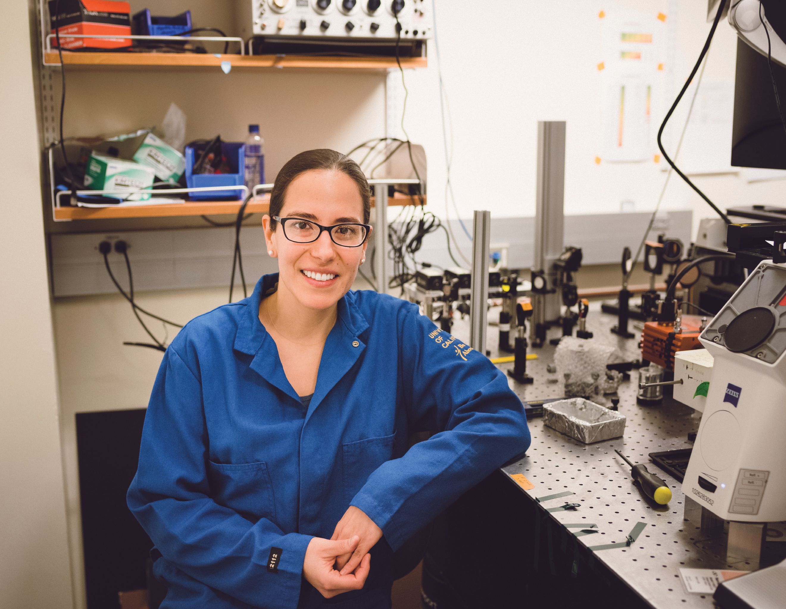
Figure 2: Landry at her laboratory optical bench on the UC Berkeley campus in 2020. (Source: Photo courtesy of Prabir Patra)

Landry is a recent recipient of 17 early career awards from the Brain and Behavior Research Foundation, the Burroughs Wellcome Fund, The Parkinson’s Disease Foundation, the Defense Advanced Research Projects Agency (DARPA) Young Investigator program, the Beckman Young Investigator program, and the Howard Hughes Medical Institute. She is also a Sloan Research Fellow and a Foundation for Food and Agriculture Research (FFAR) New Innovator.
Creating Single-Walled Carbon Nanotube-Based Sensors
Sensors developed by Landry and colleagues provide a new path for discovery of the functions of neuromodulator pathways in living cells. Daniel Heller, an associate member and associate professor at the Memorial Sloan Kettering Cancer Center–Weill Cornell Medicine at Cornell University, has known Landry since graduate school. He and Landry began collaborating on spectroscopy-related projects when they were both doctoral students, where they became friends.
“Markita has developed exciting optical nanosensors that allow the mapping of neurotransmitter release from within living neuronal tissues,” Heller said, describing her elegant approaches to develop new optical sensors for important new bioanalytical targets. “She has done much to raise the profile and excitement of this field, due to her publications that show how single-walled carbon nanotube-based sensors enable important discoveries in the fields of neuroscience and plant biology and biotechnology. She is clearly a rising superstar who has clearly chosen problems very well, tackling several areas in which she is already making a huge impact.”
Margaret Rice is a professor and the vice chair for research at the New York University (NYU) Grossman School of Medicine. She first met Landry in July 2016, when Landry was an invited speaker in the Skirball Molecular Neurobiology Seminar Series at NYU.
“She gave an outstanding talk, and I spoke with her briefly afterwards to say how impressed I was with the tools she is developing to visualize transmitters,” Rice said.
Rice later had the chance to meet with Landry and hear her speak several more times through the Parkinson’s Foundation. Landry received one of the foundation’s prestigious Fahn Fellowships in 2017, which provided three years of support for her project titled, “Direct Imaging of the Cause and Treatment of Parkinson’s Disease with Synthetic Modulatory Neurotransmitter Nanosensors.”
As a Fahn Fellow, Landry was invited to speak at the foundation’s annual Grantee Summit in 2018 and 2019.
“I am co-chairing the 2022 Basal Ganglia Gordon Research Conference, and based on Markita’s exciting work and her engaging speaking style, we invited her to be one of the speakers for this international conference,” Rice said.
Given her own extensive research studying factors that regulate dopamine release, Rice appreciates the impact of Landry’s work. Rice’s work in this area used electrochemical sensors, which record dopamine at a single site, but Landry has developed infrared dopamine nanosensors that can detect the dynamic and spatially discrete increase in brain dopamine in real time.
Figure 3: Landry sharing the fine points of her craft as a teacher and mentor at UC Berkeley in 2020. (Source: Photo courtesy of Prabir Patra)

“The methods Landry has introduced, and is continuing to refine and optimize, allow examination of multiple release sites simultaneously,” Rice said. “This will be a game changer for investigating the roles and regulation of this important neurotransmitter.”
Landry is now expanding this approach to detect other transmitters, broadening the impact of her novel methods.
Prabir Patra is the chair of Biomedical Engineering, and a professor of Biomedical Engineering and Mechanical Engineering at the School of Engineering at the University of Bridgeport, in Connecticut.
“Markita is one of the finest researchers, engineers, and collaborators that I have ever come across,” Patra said. “She is an asset to the scientific community and academia as a whole.”
Patra and Landry began working together on a project two years ago. Given their shared interest in developing nanotechnologies to advance understanding of biological systems, they undertook a project looking at polynucleotide adsorption on fluorescent graphene quantum dots. They discovered that the degree of oxidation on the surface is what determines the extent to which polynucleotides can adsorb. These findings are exciting because noncovalent conjugation of polymers to graphene quantum dots enables a new class of materials to be used for biological imaging.
“Markita has developed the first near-infrared probe to image dopamine in the brain. This is a tour-de-force body of work showing that nanomaterials can be used to image brain chemistry, which has significant implications for neuroscience in being able to study psychiatric disease,” Patra said. “Markita’s dopamine imaging probe has enabled neuroscientists to image dopamine communication at the level of individual neurons and the effects of pharmacological drugs for the first time.”
Landry’s Most Cited Research
Landry’s most cited research papers have already received more than 1200 citations, according to Google Scholar (1–8). Her most cited references involve her work as a postdoctoral researcher at the Massachusetts Institute of Technology; these are summarized below, starting with her most-cited papers.
In her most cited research, Landry and colleagues created single-walled carbon nanotubes (SWNTs) that were inserted into the lipid envelope of extracted plant chloroplasts to promote higher photosynthetic activity and maximum electron transport rates (1). In this work, SWNTs allowed NIR fluorescence monitoring of nitric oxide both ex vivo and in vivo (nitric oxide is an important physiological signaling molecule in plants), demonstrating that a plant can be altered to function as a photonic chemical sensor.
In other highly cited work, Landry used corona phase molecular recognition (CoPhMoRe) to identify adsorbed polymer phases on fluorescent single-walled carbon nanotubes (SWCNTs) for the selective detection of neurotransmitters, such as dopamine (2). This research suggests that polymer–SWCNT constructs may be useful as fluorescent neurotransmitter sensors, potentially within living tissue. Landry and coworkers have also published highly cited work on synthetic heteropolymers, constrained onto a single-walled carbon nanotube using chemical adsorption, forming a new corona phase that exhibits highly selective recognition for specific important physiologically active molecules (3). As examples, recognition complexes were developed for sensing riboflavin, L-thyroxine, and estradiol, using carbon nanotube photoemission within the NIR spectral region.
Landry has demonstrated that high-powered, NIR laser beams are useful as optical tweezers (4). Work was performed to determine the potential damage caused by using these optical traps or “tweezers” for DNA molecules tethered between optically trapped polystyrene microspheres. The use of optical tweezers is important for the study of molecules using laser-based spectroscopic methods. This work is important due to the most recent involvement of the study of DNA as an analyte molecule.
Landry’s work on carbon nanomaterials, also highly cited, has been shown to be useful in biomedicine for imaging, sensing, targeting, delivery, therapeutics, catalysis, and energy harvesting (5). The carbon scaffold and a selected biological moiety are used to form a complex interface; such an interface presents a physiochemical challenge for achieving efficient and robust binding, while preserving biological activity of molecules. There is important discovery research still required to understand carbon nanomaterials as protein carriers in order to maintain the molecular performance and activity of the biological moieties. Landry’s team has performed research demonstrating these concepts and is moving closer to discovering basic functionality and further applications.
Nanosensor arrays are known to provide ultralow detection limits in reagent solutions when an analyte is in close proximity (6). In Landry’s work in this area, label-free detection arrays were demonstrated to be capable of measuring individual proteins from Escherichia coli (bacteria) and Pichia pastoris (yeast) that were immobilized within a microfluidic chamber, enabling the measurement of protein efflux from single organisms in real time. These sensor arrays are fabricated using non-covalent conjugation of an aptamer-anchor polynucleotide sequence to NIR fluorescence emissive single-walled carbon nanotubes. Such a sensor produces a fluorescence signal when activated by the analyte species.
Corona phase molecular recognition (CoPhMoRe) consists of a heteropolymer adsorbed onto a nanoparticle surface to recognize a specific target analyte, such as fibrinogen (7). Landry and her coworkers developed a variant of a CoPhMoRe screening procedure using single-walled carbon nanotubes (SWCNTs), and tested it against a panel of human blood proteins, revealing a specific corona phase that recognizes fibrinogen with high selectivity and good sensitivity.
In other work by Landry and colleagues, SWCNTs allowed the development of ratiometric fluorescent sensors for the first time (8). Single chirality SWCNTs were independently functionalized to detect either nitric oxide (NO), (a transient cellular signaling molecule), hydrogen peroxide (H2O2) (another known biological signaling molecule), or no analyte (remaining invariant), to create optical sensor responses from the ratio of distinct fluorescent emission peaks. This SWCNT ratiometric sensing platform is providing the detection of trace analytes in strongly scattering media, such as found in biological tissues. Such sensors are unique and noteworthy in biomedical research.
Recent Research
Landry’s most recent research has demonstrated that specially covalently functionalized SWCNTs will maintain their intrinsic fluorescence-based molecular recognition for selected neurotransmitter and protein analytes (9). Her group discovered that a targeting ligand, such as biotin, allowed the SWCNTs to self-assemble on passivated microscopy slides for future use in multiplexed analyte sensing and imaging applications. This work is leading to the development of robust biomolecule sensors.
Landry and her coworkers have recently demonstrated time-resolved measurements of exchange dynamics between solution-phase and adsorbed corona-phase DNA and protein biomolecules on SWCNTs to study protein corona dynamics on ssDNA-SWCNT–based dopamine sensors (10).
In other current work, a responsive and reversible generic platform was developed to provide synthetic molecular recognition of the neuromodulator serotonin on the surface of NIR fluorescent SWCNT signal transducers (11). The results suggest that such nanosensors could be developed for other important neuromodulators in tissue, to study the fundamental aspects of neurotransmission.
Her exciting work in genetic engineering is described in papers published last year (12,13). In these papers, Landry’s group has demonstrated the application of nanomaterial variants and associated surface chemistries to controllably graft and quickly deliver functional “biomolecular cargo,” such as whole DNA plasmids, into various agriculturally important plants.
“We accomplish nanoparticle-mediated heterologous and transient expression of a protein in plants, and show that there is no transgenic DNA integration into the host plant genome,” Landry explains.
This research has been demonstrated to enable gene expression in a series of agriculturally meaningful plants, including Nicotiana benthamiana (tobacco, a model dicot plant), Eruca sativa (arugula, a non-model dicot plant); Triticum aestivum (wheat, a non-model monocot plant), and Gossypium hirsutum (cotton, a non-model dicot plant).
“Wheat and cotton are cash crops; cotton, in particular, are challenging to genetically transform with current technologies,” she notes. “We further demonstrate with single-molecule imaging that the process of grafting DNA on nanoparticles prevents DNA degradation by nucleases, providing a complete picture of how this nanotechnology both promotes delivery of functional biomolecules to plants, and also protects cargo from enzymatic degradation en route to its intracellular function.”
Exceptional Teacher and Mentor
Landry spends significant time and effort teaching, mentoring, and carefully developing her graduate students’ research and scientific maturity.
“In addition to the impressive scholarship produced by Dr. Landry’s laboratory, so early in her career her lab has already produced several faculty members at top institutions,” said Heller. “She is an exceptional mentor and inspiration to
young scientists.”
Patra agrees,.“In addition to her science, I think it is impressive that the graduates from Markita’s lab are achieving exceptional success in their careers,” he said.
As examples, he notes that Landry’s former student leading the brain imaging work is now a professor at the Howard Hughes Medical Institute (HHMI) Janelia Research campus, that another former student who was leading nanosensor development is now a scientist at Gordian Biotechnology, and a former student who was leading plant genetic transformation work will soon be a professor at Caltech.
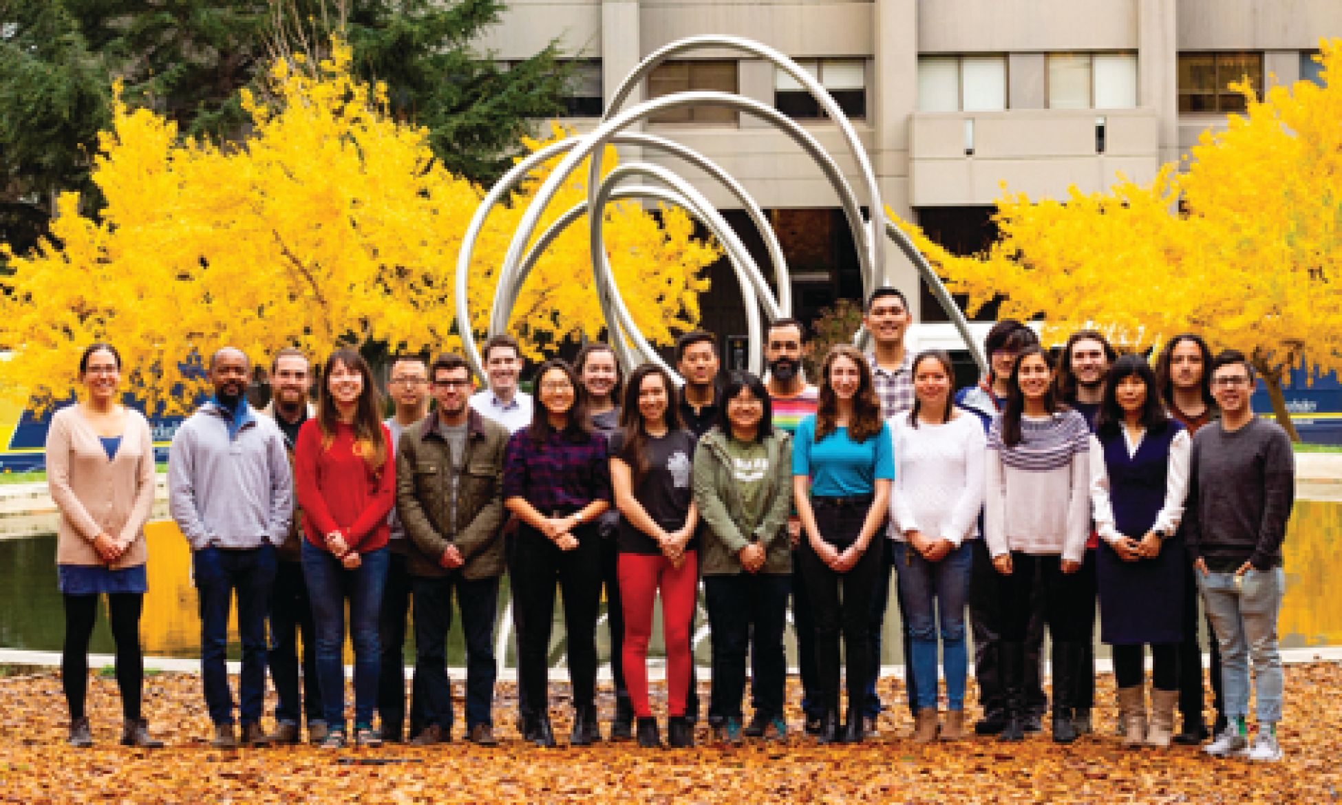
Figure 4: Landry’s UC Berkeley group from 2019, from left to right: Markita Landry, Abraham Beyene, Gabriel Dorlhiac, Alison Lui, Sanghwa Jeong, Nicholas Ouassil, Ian McFarlane, Sarah Yang, Nicole Navarro, Madeline Klinger, Darwin Yang, Natalie Goh, Eduardo Gonzalez Grandio, Rebecca Pinals, Francis Ledesma, Linda Chio, Jeffrey Wang, Gozde Demirer, Josh Hubbard, Huan Zhang, Frankie Cunningham, Chris Jackson. (Source: Photo courtesy of M. Landry)

The Future
Complex research involving the development of nanosensors is a fascinating field, and Landry appears poised to continue to make important contributions in this area. “I have watched with awe as Dr. Landry’s academic career has taken off due to her impressive scholarship in the field of nanotechnology, specifically in the area of single-walled carbon nanotube-based technologies,” Heller said. “I think Markita’s career will lead to a bright future.”
Rice agrees. “Markita is already a leader in the field—indicated by her being a sought-after speaker at international meetings,” she said. “I have no doubt this upward trajectory will continue as she continues to pioneer novel detection methods for important molecules.”
Patra also looks forward to Landry’s continued research. “Markita is an early career researcher but has already established herself as a world leader in the field of nanotechnology, neuroscience, and agriculture,” he said. “The science that has come out of the Landry lab since its start four years ago is nothing short of extraordinary, so wherever she chooses to take her career next I’m sure will be at least as exciting.”
References
- J.P. Giraldo, M.P. Landry, S.M. Faltermeier, T.P. McNicholas, N.M. Iverson, A.A Boghossian, N.F. Reuel, A.J. Hilmer, F. Sen, J.A. Brew, and M.S. Strano, Nat. Mater. 13(4), 400–408 (2014).
- S. Kruss, M.P. Landry, E. Vander Ende, B.M. Lima, N.F. Reuel, J. Zhang, J. Nelson, B. Mu, A. Hilmer, and M. Strano, J. Am. Chem. Soc. 136(2), 713–724 (2014).
- J. Zhang, M.P. Landry, P.W. Barone, J.H. Kim, S. Lin, Z.W. Ulissi, D. Lin, B. Mu, A.A. Boghossian, A.J. Hilmer, A. Rwei, A.C. Hinckley, S. Kruss, M.A. Shandell, N. Nair, S. Blake, F. Şen, S. Şen, R.G. Croy, D. Li, K. Yum, J.H. Ahn, H. Jin, D.A. Heller, J.M. Essigmann, D. Blankschtein, and M.S. Strano, Nat. Nanotechnol. 8(12), 959–968 (2013).
- M.P. Landry, P.M. McCall, Z. Qi, and Y.R. Chemla, Biophys. J. 97(8), 2128–2136 (2009).
- S.F. Oliveira, G. Bisker, N.A. Bakh, S.L. Gibbs, M.P. Landry, M.S. Strano, Carbon 95, 767–779 (2015).
- M.P. Landry, H. Ando, A.Y. Chen, J. Cao, V.I. Kottadiel, L. Chio, D. Yang, J. Dong, T.K. Lu, and M.S. Strano, Nat. Commun. 12(4), 368 (2017).
- G. Bisker, J. Dong, H.D. Park, N.M. Iverson, J. Ahn, J.T. Nelson, M.P. Landry, S. Kruss, and M.S. Strano, Nat. Commun. 7(1), 1–14 (2016).
- J.P. Giraldo, M.P. Landry, S.-Y. Kwak, R.M. Jain, M.H. Wong, N.M. Iverson, M. Ben-Naim, M.S. Strano, Small. 11(32), 3973–3984 (2015).
- L. Chio, R.L. Pinals, A. Murali, N.S. Goh, and M.P. Landry, Adv. Funct. Mater. 30(17), 1910556 (2020).
- R.L. Pinals, D. Yang, A. Lui, W. Cao, and M.P. Landry, J. Am. Chem. Soc. 142(3), 1254–1264 (2020).
- S. Jeong, D.Yang, A.G. Beyene, J. Travis Del Bonis-O’Donnell, A.M.M. Gest, N. Navarro, X. Sun, and M.P. Landry, Sci. Adv. 5(12), 37–71 (2019).
- G.S. Demirer, H. Zhang, J. Matos, N. Goh, F.J. Cunningham, Y. Sung, R. Chang, A.J. Aditham, L. Chio, M.J. Cho, B. Staskawicz, and M.P. Landry, Nat. Nanotechnol. 14, 456–464 (2019).
- H. Zhang, G.S. Demirer, H. Zhang, T. Ye, N.S. Goh, A.J Aditham, F.J. Cunningham, C. Fan, and M.P. Landry, Proc. Natl. Acad. Sci. U.S.A. 116(15), 7543–7548 (2019). DOI: 10.1073/pnas.1818290116.
Jerome Workman, Jr. is the Senior Technical Editor of Spectroscopy. Direct correspondence to jworkman@mjhlifesciences.com.

Multi-Analytical Study Reveals Complex History Behind Ancient Snake Motif in Argentine Rock Art
May 22nd 2025A recent study published in the Journal of Archaeological Science: Reports reveals that a multi-headed snake motif at Argentina's La Candelaria rock shelter was created through multiple painting events over time.
Chinese Researchers Develop Dual-Channel Probe for Biothiol Detection
April 28th 2025Researchers at Qiqihar Medical University have developed a dual-channel fluorescent probe, PYL-NBD, that enables highly sensitive, rapid, and selective detection of biothiols in food, pharmaceuticals, and living organisms.
New Study Reveals Insights into Phenol’s Behavior in Ice
April 16th 2025A new study published in Spectrochimica Acta Part A by Dominik Heger and colleagues at Masaryk University reveals that phenol's photophysical properties change significantly when frozen, potentially enabling its breakdown by sunlight in icy environments.

.png&w=3840&q=75)

.png&w=3840&q=75)


.png&w=3840&q=75)


.png&w=3840&q=75)


