The Infrared Spectra of Polymers, Part I: Introduction
Polymers are perhaps the most common type of sample analyzed by infrared spectroscopy. In this first of a series of columns, I will introduce the properties of polymers, discuss what infrared spectroscopy can tell us about polymers, and begin the analysis of the spectra of polymers with a look at the spectrum of polyethylene.
I have over 30 years experience as an infrared (IR) spectroscopist, and have written three books on the subject (1–3). I have also taught infrared spectral interpretation to thousands of people as part of the training courses I offer. In my experience, the sample type most frequently analyzed by IR is polymers. Since polymers are so important, why have I waited until the seventh year of this column to discuss them?
What’s unique about polymers is that they contain practically every functional group imaginable, so we weren’t ready to cover polymers until we had discussed all the possible functional groups they contain, and, yes, that has taken several years. Now that we have a solid foundation, it is time to look at the spectra of polymers in detail.
Introduction to Polymers
What makes polymers unique compared to other molecules is that they are comprised of a series of identical chemical structures that form a chain. Each link in the chain is called a repeat unit. The word polymer is derived from the Greek words “poly,” or many, and “mer,” or parts. Thus, a polymer is a molecule that contains many parts. Polymers are synthesized from small molecules called monomers, Greek for “one part.” Polymers are named by appending the prefix “poly-” to the name of the monomer. The chemical structure of a common polymer, polyethylene, is seen in Figure 1.
FIGURE 1: The chemical structure of polyethylene.

Polyethylene is named by appending “poly-” to the name of the monomer “ethylene.” The repeat unit in polyethylene is the CH2 or methylene group. Note from Figure 1 that the chemical structure of a polymer is drawn with brackets around the repeat unit and a subscript n. The n means there are some number n of repeat units in a row comprising the polymer chain. Note that there are dangling chemical bonds to the left and right of the repeat unit denoting bonds to repeat units to the left and right.
For small molecules, we are used to thinking of the molecular weight as a constant. For example, the molecular weight of every molecule of CO2 in the universe (assuming the atomic mass of carbon = 12 and that of oxygen = 16) is 44. This is not true of polymers. The complication is that almost all polymer samples consist of molecules with different numbers of repeat units, and therefore different molecular weights. For example, if we assume the atomic mass of the repeat unit in polyethylene is 14, a chain with 100 repeat units will have a molecular weight of 1400, while a molecule containing 1000 repeat units will have a molecular weight of 14,000. Polymer chains with molecular weights upwards of a million are known (4).
The challenge, then, is if we have chains of different molecular weight in a sample, what number do we use to characterize the molecular weight? Rather than speaking of a specific molecular weight, we measure and use what is called a molecular weight distribution (4). An example of a possible molecular weight distribution curve for a polymer sample is seen in Figure 2.
FIGURE 2: A hypothetical molecular weight distribution of a polymer sample. Note that the x-axis units are molecular weight, and the y-axis units are number of molecules.
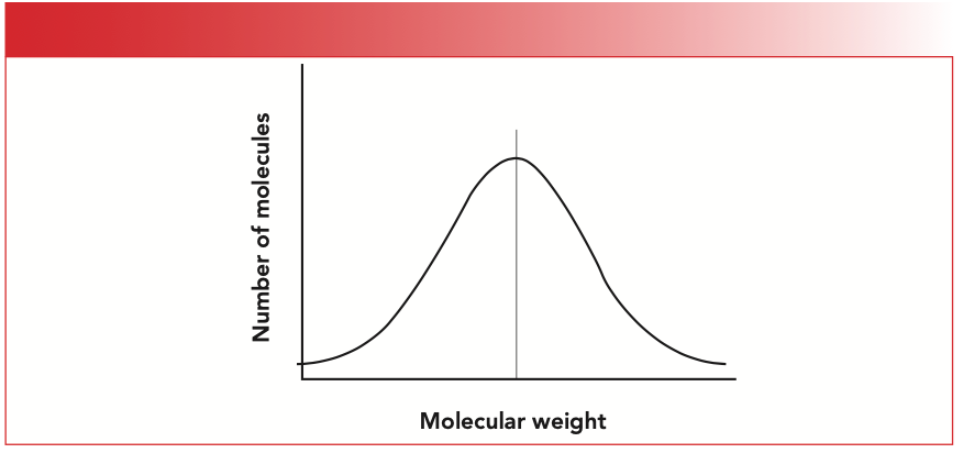
Note that the figure is a plot of the number of molecules versus molecular weight. There is a lot of information to be derived from plots like the one in Figure 2, and various experimental methods of measuring them, all of which are beyond the scope of this article (4).
So, if the molecular weight distribution of a polymer sample is important, what can infrared spectroscopy tell us about molecular weight distribution? Absolutely nothing! Remember that infrared spectra contain functional group structural information (2). In a sample of polyethylene, each and every repeat unit is structurally identical (ignoring end groups), so each and every repeat unit will therefore have the same spectrum. For example, the spectrum of a polyethylene molecule with 100 repeat units will be the same for all practical purposes to the spectrum of a polyethylene molecule that contains 1000 repeat units. Infrared spectroscopy tells us the structure of polymeric repeat units; it tells us little about molecular weight distributions.
What, then, should we expect polymer spectra to look like? Recall that the number of vibrations for a non-linear molecule is 3N-6, where N is the number of atoms in the molecule (2). Also, recall that the number of vibrations in a molecule is determined in part by the number of vibrations it possesses and the symmetry of the molecule (2). A polyethylene molecule has 4 atoms in each repeat unit, and if there are 1000 repeat units in a chain, that means there are 4000 atoms present. This molecule then has (3 x 4000) - 6), or 11,994, vibrations, and, in principle, should have a complex spectrum with perhaps thousands of peaks.
In fact, nothing could be further from the truth. The infrared spectrum of a polyethylene sample is seen in Figure 3.
FIGURE 3: The infrared spectrum of a low density polyethylene (LDPE) sample. Note the small number of peaks.
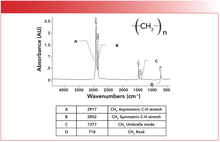
Note that there are not thousands of peaks in this spectrum, but a handful. This is because, although there are many atoms and many vibrations for polyethylene molecules, the molecule has high symmetry, because each and every repeat unit is structurally the same, and hence will give the same spectrum. The complexity of a polymer spectrum then is determined not so much by the number of atoms total in the chain, but by the number of atoms in the repeat unit. This is why the spectrum of polyethylene is so simple even though it has a high molecular weight.
The Infrared Spectroscopy of Polyethylene
Given the simplicity of Figure 3, you may think that there is not much to learn from the spectra of this polymer, but the subtleties and complexities of its spectrum may surprise you. Since polymers contain functional groups whose spectra we have already covered, we know everything we need to know to interpret polymer spectra. For example, the CH2 group in polyethylene is very similar to the methylene groups in molecules like hexane and octane whose spectra we have already examined (5,6), so all the rules we have learned about how to interpret the spectra of methylene groups apply to polyethylene as well.
Table I summarizes the group wavenumbers for CH2 groups. Using Table I, we can easily analyze the spectrum Figure 1. The peak labeled A at 2917 cm-1 is the methylene asymmetric stretch, the peak labeled B at 2852 is the symmetric stretch, and the peak labeled D at 718 is the rock. Recall that the 720 peak only appears if there are four or more methylenes in a row (6), which is true of probably every polyethylene molecule.

Recall that the number of CH stretching peaks between 3000 and 2850 can be used to tell us whether a molecule contains methyls only, methylenes only, or both (6). This info is summarized in Table II.
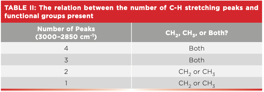
Polyethylene clearly has methylenes only, since it has two peaks between 3000 and 2850.
Polyethylene: More Than Meets the Eye
Note in Figure 3 that there is a peak labeled C at 1377 assigned as a methyl group (CH3) umbrella mode. What’s up with that? Haven’t we said that polyethylene contains CH2 groups only, as seen by the number of C-H stretching peaks? Yes, but Mother Nature does love to throw us curve balls sometimes. Because of the specific chemical process used to make the sample of polyethylene whose spectrum is seen in Figure 3, there are a small number of repeat units that have what are called side chains; these are short alkyl chains whose structures include –(CH2)5- CH3. An example of a side chain is seen in Figure 4.
FIGURE 4: An example of an alkyl side chain present in some polyethylene molecules.
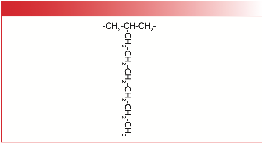
Polyethylene made via this reaction is called low density polyethylene (LDPE) because the alkyl side chains push the polymer chains apart, reducing the density of the material. As a result, LDPE is structurally weak, and is used to make things like plastic bags that can be easily torn apart with bare hands. The methyl groups in the side chains are responsible for the umbrella mode peak at 1377. The C-H stretches from these groups are swamped by the CH2 stretching peaks, so we do not see them.
So, LDPE is not really “pure” polyethylene because of the presence of some methyl groups. Is there a polyethylene sample devoid of side chains, and contains CH2s only? Yes, using a different chemical reaction, side-chain-free polyethylene can be made. This material is called high density polyethylene (HDPE), and its infrared spectrum is seen in Figure 5.
FIGURE 5: The infrared spectrum of high density polyethylene (HDPE).
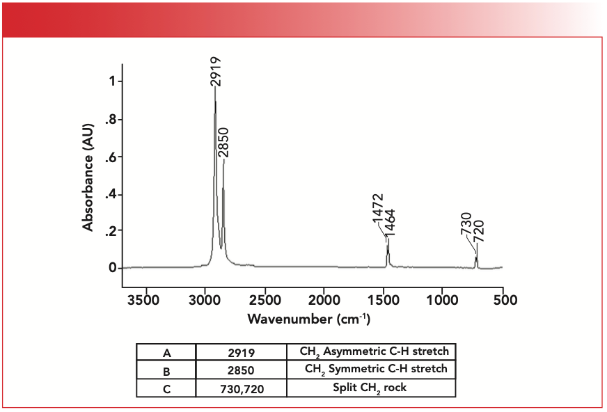
The spectrum of HDPE has the methylene stretching and rocking peaks as expected, and does not have a methyl group umbrella mode, because there are no CH3 groups present because of the absence of side chains. HDPE is high density because the polymer chains can pack close together (thanks to the absence of side chains), giving a denser material. As a result, HDPE is structurally stronger than LDPE, and is made into things like milk jugs, which are difficult to tear apart with bare hands. The easiest way to distinguish between LDPE and HDPE is that spectra of LDPE have a CH3 umbrella mode, while spectra of HDPE do not.
What’s with the split rocking mode peak in Figure 5 at 730/720? More on that and the fascinating spectra of polymers next time.
Conclusions
The infrared spectra of polymers do not measure molecular weight distributions, but the structure of the repeat units in polymers. The spectra of functional groups in polymers are similar to those in small molecules. The spectra of low-density and high-density polyethylene were discussed, showing some of the nuances of polymer spectra.
References
(1) B.C. Smith, Fundamental of Fourier Transform Infrared Spectroscopy (CRC Press, Boca Raton, Florida, 2nd Ed., 2011).
(2) B.C. Smith, Infrared Spectral Interpretation: A Systematic Approach (CRC Press, Boca Raton, Florida, 1999).
(3) B.C. Smith, Quantitative Spectroscopy: Theory and Practice (Elsevier, New York, New York, 2002).
(4) F. Bovey and F.H. Winslow, Macromolecules: An Introduction to Polymer Science (Academic Press, New York, New York, 1980).
(5) B.C. Smith, Spectroscopy 30(4), 18–23 (2015).
(6) B.C. Smith, Spectroscopy 30(7), 26– 31, 48 (2015).
Brian C. Smith, PhD, is the founder and CEO of Big Sur Scientific, a maker of portable mid-infrared cannabis analyzers. He has over 30 years experience as an industrial infrared spectroscopist, has published numerous peer-reviewed papers, and has written three books on spectroscopy. As a trainer, he has helped thousands of people around the world improve their infrared analyses. In addition to writing for Spectroscopy, Dr. Smith writes a regular column for its sister publication Cannabis Science and Technology and sits on its editorial board. He earned his PhD in physical chemistry from Dartmouth College. He can be reached at: SpectroscopyEdit@MMHGroup.com ●


AI Shakes Up Spectroscopy as New Tools Reveal the Secret Life of Molecules
April 14th 2025A leading-edge review led by researchers at Oak Ridge National Laboratory and MIT explores how artificial intelligence is revolutionizing the study of molecular vibrations and phonon dynamics. From infrared and Raman spectroscopy to neutron and X-ray scattering, AI is transforming how scientists interpret vibrational spectra and predict material behaviors.
Real-Time Battery Health Tracking Using Fiber-Optic Sensors
April 9th 2025A new study by researchers from Palo Alto Research Center (PARC, a Xerox Company) and LG Chem Power presents a novel method for real-time battery monitoring using embedded fiber-optic sensors. This approach enhances state-of-charge (SOC) and state-of-health (SOH) estimations, potentially improving the efficiency and lifespan of lithium-ion batteries in electric vehicles (xEVs).
New Study Provides Insights into Chiral Smectic Phases
March 31st 2025Researchers from the Institute of Nuclear Physics Polish Academy of Sciences have unveiled new insights into the molecular arrangement of the 7HH6 compound’s smectic phases using X-ray diffraction (XRD) and infrared (IR) spectroscopy.