The Infrared Spectra of Polymers, VI: Polymers With C-O Bonds
This column series has been organized into sections based on the infrared (IR) spectroscopy of specific chemical bonds. We have discussed functional groups containing C-H, C-O, and C=O bonds. Here, in the sixth installment of our survey of the IR spectra of polymers, we discuss polymers that contain C-O bonds. The spectra we will examine include polyvinyl alcohol, cellulose, starch, and copolymers that contain C-O bonds such as polyethetherketone, polyurethanes, and polyimides. The unifying theme of all these spectra is that they all contain one or more large C-O stretching peaks between 1300–1000 cm-1.
We covered the spectroscopy of the C-O bond in earlier columns (1–4). In those installments, the functional groups we studied were alcohols and ethers. Recall that the overriding spectral feature of C-O bonds are one or more large C-O stretching peaks between 1300–1000 cm-1 (1) (going forward, assume all peak positions are in cm-1 units unless otherwise noted). Usually, the C-O stretch is the most intense peak in this wavenumber region.
Recall that alcohols hydrogen bond strongly to each other similar to water (1,2). An example of alcohols hydrogen bonding is seen in Figure 1.
FIGURE 1: An example of two alcohol molecules hydrogen bonding to each other.

The effect of hydrogen bonding on the spectra of alcohols is that any peaks from O-H stretching and bending motions are unusually broadened, which is illustrated in the spectrum of ethanol seen in Figure 2.
FIGURE 2: The mid-infrared (mid-IR) spectrum of ethanol (ethyl alcohol). Note that peaks A and E are broadened compared to the rest of the peaks in the spectrum.
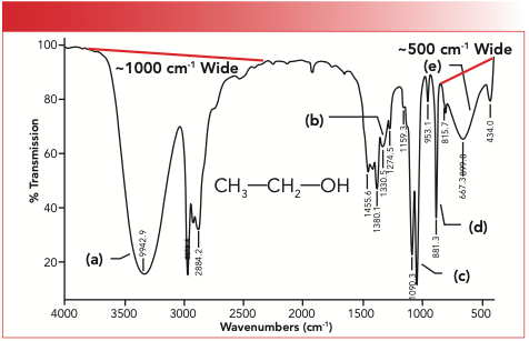
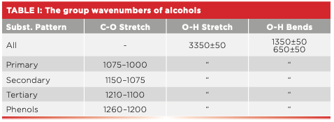
Note in the figure that the O-H stretching peak labeled A at 3492 is approximately 1000 cm-1 wide at its base, and the O-H wagging peak labeled E at 667 is approximately 500 cm-1 wide at its base. These peaks are significantly broader than the peaks from other functional groups seen in this spectrum. In general, the O-H stretching peaks of alcohols fall at 3350±50, whereas the O-H wag is normally seen at 650±50. The C-O stretching feature of alcohols falls from 1260 to 1000, with the range being determined as to whether the alcohol is primary, secondary, tertiary, or a phenol. The C-O stretching peak of ethyl alcohol, a primary alcohol, is labeled C at 1050, which is in the range expected. A summary of the group wavenumbers for alcohols is seen in Table I.
In addition to alcohols, another functional group containing C-O bonds found in polymers is ethers. The chemical structure of ethers is characterized by two C-O bonds, which is why two ether carbons can be seen in Figure 3.
FIGURE 3: The molecular structure of the ether functional group.

If both ether carbons are saturated, the ether is said to be saturated. If both ether carbons are aromatic, the ether is aromatic. And finally, if one ether carbon is saturated and the other is aromatic, we have a mixed ether. Note that ethers do not contain O-H bonds. Therefore, they do not hydrogen bond, do not contain broadened peaks, and do not have O-H stretching and bending vibrations like alcohols. In fact, the only useful group wavenumber that ethers have is a C-O stretch, which is typically one or two peaks that fall from 1300 to 1070. A summary of the group wavenumbers for ethers is seen in Table II.

Note in Table II that saturated ethers have one C-O stretching peak from 1140–1070, aromatic ethers have one peak from 1300–1200, and mixed ethers have two C-O stretching peaks—one at 1050–1010 and the other at 1300–1200. The reason mixed ethers have two C-O stretching peaks is that the two ether carbons are bonded to different types of carbon so are chemically different, have different C-O stretching force constants, and hence have different peak positions (5). For mixed ethers then, the aromatic C-O stretching vibration gives the peak from 1300 to 1200, whereas the saturated ether C-O stretching peak falls from 1050 to 1010. Note that for both mixed and aromatic ethers, the aromatic C-O stretch has the same range of 1300–1200. As a general rule, if an ether exhibits a C-O stretching peak from 1300 to 1200, then at least one of the ether carbons is aromatic.
The Infrared (IR) Spectra of O-H Containing Polymers
Polyvinyl Alcohol
The way in which polyvinyl alcohol is synthesized is unusual in that rather than being made from an O-H containing monomer, one of the reactants is a polymer. This reaction is shown in Figure 4.
FIGURE 4: The reaction via which polyvinyl alcohol is made from polyvinyl acetate.
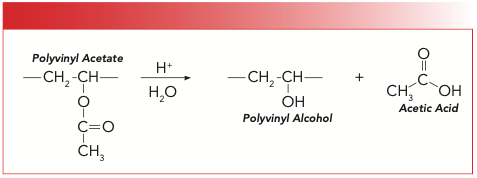
The reactant is polyvinyl acetate, which contains a pendant acetate moiety, which is an ester. Polyvinyl acetate is hydrolyzed with aqueous acid to give polyvinyl alcohol while splitting off a molecule of acetic acid. The IR spectrum of polyvinyl alcohol is seen in Figure 5.
FIGURE 5: The IR spectrum of polyvinyl alcohol.
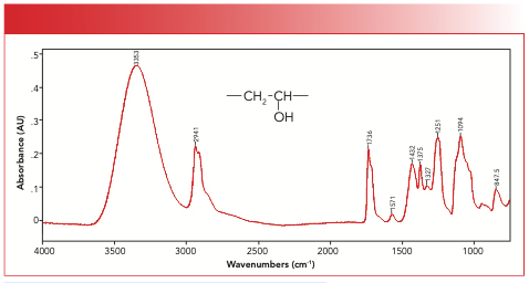
We have the classic broad, intense O-H stretch at 3353, and the C-O stretch of a secondary alcohol at 1094. This particular spectrum cuts off at approximately 700, so the expected O-H wag at ~650 is not visible. Another thing to notice in Figure 5 is the presence of peaks from unreacted acetate, giving us the classic ester “Rule of 3” peaks (6) at 1736, 1251, and 1094. Note how the peak at 1094 is broadened. My interpretation of this peak is that it is an overlap of the alcohol and ester C-O stretching peaks. Despite the ester peaks, polyvinyl alcohol is easily identifiable because it contains an alcohol group, courtesy of the broadened peaks at 3353 and 1094.
The IR Spectra of Polymers Containing Ether Linkages
Polyacetal
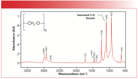
The polyacetal molecule is made by polymerizing formaldehyde, which has the formula H2-C=O. The C=O bond in formaldehyde is opened up, forming polyacetal, whose structure and spectrum are seen in Figure 6. Note the C-O stretching peak of polyacetal at 1090, as expected for a saturated ether.
FIGURE 6: The structure and spectrum of polyacetal. Note the saturated ether C-O stretching peak at 1090.
Polyetheretherketone (PEEK)
The fact that the name of this polymer contains the word ether twice just might make you think that it contains ether linkages, which it does. This material is, in fact, a copolymer made from monomers that form ketone and ether linkages upon the polymer’s formation. We will talk about polymers containing ketones in a future column. The structure and spectrum of PEEK are shown in Figure 7.
FIGURE 7: The IR spectrum of polyetheretherketone (PEEK). Note the aromatic C-O stretch at 1227.
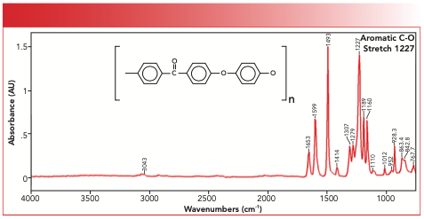
This structure contains aromatic ether linkages only, and the aromatic C-O stretching peak falls at 1227 as expected.
Foam Rubber
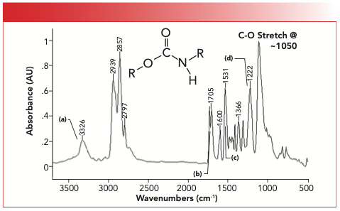
Foam rubber, the bouncy stuff that we often sit and jump on, contains a urethane linkage whose structure is seen in Figure 8 (we will cover polyurethanes in a future column). However, the urethane is often copolymerized with a monomer that gives a saturated ether linkage. We know this because the spectrum of a foam rubber sample seen in Figure 8 has a C-O stretching peak at 1050, consistent with the presence of a saturated ether linkage.
FIGURE 8: The IR spectrum of a sample of foam rubber. Although the urethane peaks dominate the spectrum, there is a C-O stretching peak from ether linkages at 1050.
Kapton
Kapton is the trade name for an engineering polymer that contains the imide functional group, which we will cover in a future column. However, note from the structure of Kapton in Figure 9 that it contains aromatic ether linkages as well.
FIGURE 9: The IR spectrum of Kapton, an imide-ether copolymer. Note the C-O stretching peak at 1241.
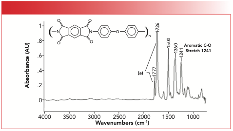
Note that the spectrum of Kapton contains a peak at 1241 assigned as the aromatic C-O stretching peak of its aromatic ether linkages. This peak is in the middle of the range where it should fall, which is from 1300 to 1200 cm-1.
Polymers that Contain Both Alcohol and Ether Groups
Carbohydrates and Polysaccharides
Carbohydrates are biological molecules that contain both alcohol and ether link- ages, which means their C-O stretching regions are complex. The building blocks of carbohydrates are sugar units or monosaccharides. When we build a polymer out of these sugar units, we get a polysaccharide. We covered the spectra of carbohydrates in a previous column (4).
A common polysaccharide is cellulose, which makes up the cell walls of plants and is otherwise known as wood, paper, or cotton. Cellulose is a high polymer of the monosaccharide glucose—the product of photosynthesis. The structure and IR spectrum of cellulose are seen in Figure 10.
FIGURE 10: The infrared spectrum of cellulose, a polysaccharide.
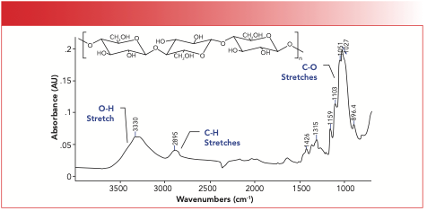
The glucose units in cellulose are stitched together by saturated ether linkages. There is so much hydrogen bonding in cellulose that the complex -O-H stretching peak is a broad blob. The C-H stretches are similarly blobby, and the C-O stretching region is best characterized as an envelope with any number of shoulder peaks on top of it.
Another important and well-known polysaccharide is starch. Starch is also a high polymer of glucose, but the saturated ether linkages that connect the glucose molecules in starch have a different orientation than in cellulose, which can be seen by comparing the structure of cellulose in Figure 10 to the structure of starch in Figure 11. Figure 11 also shows the IR spectrum of starch.
FIGURE 11: The IR spectrum of starch.
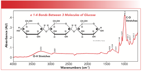
One of the maxims about IR spectroscopy is that if two molecules have different chemical structures, they will have different IR spectra (5). Cellulose and starch are similar but not identical molecules; therefore, they have similar but not identical IR spectra. For example, note that the O-H stretching envelopes of cellulose and starch top out at 3350 and 3258, respectively, and that their complex C-O stretching envelopes peak out at 1027 and 998, respectively. Additionally, the shoulders on the C-O stretching envelope in both spectra are at distinct positions, which means that one can readily distinguish between cellulose and starch via IR spectroscopy.
Conclusions
This article was devoted to polymers containing C-O bonds that are not epoxies. We reviewed the spectroscopy and group wavenumbers of alcohols and ethers, two significant functional groups that contain C-O bonds. We examined the spectrum of polyvinyl alcohol, found that its O-H stretching and bending peaks are broadened because of hydrogen bonding, and that its C-O stretching peaks falls as expected. Then, we looked at the spectra of polymers containing ether linkages, and examined the spectra of polyacetal, polyetheretherketone, urethane foam, and Kapton. Lastly, we examined the spectra of polysaccharides that contain both alcohol and ether groups and found that we can use IR spectroscopy to distinguish cellulose from starch.
References
(1) B.C. Smith, Spectroscopy 32(1), 14–21 (2017).
(2) B.C. Smith, Spectroscopy 32(4), 19–23 (2017).
(3) B.C. Smith, Spectroscopy 32(5), 22–26 (2017).
(4) B.C. Smith, Spectroscopy 32(7), 18–24 (2017).
(5) B.C. Smith, Spectroscopy 30(1), 16–23 (2015).
(6) B.C. Smith, Spectroscopy 33(7), 20–23 (2018).
Brian C. Smith, PhD, is the founder and CEO of Big Sur Scientific, a maker of portable mid-infrared cannabis analyzers. He has over 30 years experience as an industrial infrared spectroscopist, has published numerous peer-reviewed papers, and has written three books on spectroscopy. As a trainer, he has helped thousands of people around the world improve their infrared analyses. In addition to writing for Spectroscopy, Dr. Smith writes a regular column for its sister publication Cannabis Science and Technology and sits on its editorial board. He earned his PhD in physical chemistry from Dartmouth College. He can be reached at: SpectroscopyEdit@MMHGroup.com ●


AI Shakes Up Spectroscopy as New Tools Reveal the Secret Life of Molecules
April 14th 2025A leading-edge review led by researchers at Oak Ridge National Laboratory and MIT explores how artificial intelligence is revolutionizing the study of molecular vibrations and phonon dynamics. From infrared and Raman spectroscopy to neutron and X-ray scattering, AI is transforming how scientists interpret vibrational spectra and predict material behaviors.
Real-Time Battery Health Tracking Using Fiber-Optic Sensors
April 9th 2025A new study by researchers from Palo Alto Research Center (PARC, a Xerox Company) and LG Chem Power presents a novel method for real-time battery monitoring using embedded fiber-optic sensors. This approach enhances state-of-charge (SOC) and state-of-health (SOH) estimations, potentially improving the efficiency and lifespan of lithium-ion batteries in electric vehicles (xEVs).
New Study Provides Insights into Chiral Smectic Phases
March 31st 2025Researchers from the Institute of Nuclear Physics Polish Academy of Sciences have unveiled new insights into the molecular arrangement of the 7HH6 compound’s smectic phases using X-ray diffraction (XRD) and infrared (IR) spectroscopy.