Applications of 1064 nm Dispersive Raman Systems in Biofuel Research
BaySpec, Inc. has developed a complete line of 1064 nm excitation, dispersive Raman systems that offer maximum reduction in fluorescence interference from biological samples and thus making them very useful tools for biofuel research.
Jason Hammock, Peijun Cong, Eric Bergles, and William Yang, BaySpec, Inc.
BaySpec, Inc. has developed a complete line of 1064 nm excitation, dispersive Raman systems that offer maximum reduction in fluorescence interference from biological samples and thus making them very useful tools for biofuel research.
Biofuels are a current focus of government funding (1) and research efforts from academia, as well as industry, as we look to reduce green house gas emissions and secure our energy future away from fossil fuels. Of these materials being evaluated for biofuel production, algae offers the single greatest promise of becoming a sustainable source as our transportation fuel and at the same time does not produce net green house gas emissions in its life cycle. Algae are unique organisms that can store energy in the form of lipids which can comprise up to 60% of their body mass. These lipids can be readily converted to transportation fuel, such as biodiesel. As different micro and macro algae are evaluated, timely quantitation of their lipid content becomes increasingly important.
Traditional, wet chemistry-based methods are time consuming, require large amounts of sample, and are tedious to implement. Fluorescence based methods allow in-situ lipid content measurement, however, the algae samples have to be stained first with fluorescence labeling molecules (2). Raman spectroscopy is a non-destructive technique that measures the vibrational "fingerprint" and requires essentially no sample preparation and, thus, would be ideal for these applications. Unfortunately, strong background fluorescence from algae tends to overwhelm the Raman spectral features, even with 785 nm laser excitation.
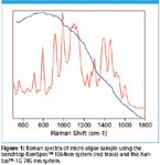
BaySpec's line of 1064 nm Raman analyzers offer the means to minimize interference from fluorescence and unmask much of the algae Raman spectral "fingerprint." In Figure 1, a sample of micro-algae has been analyzed using the benchtop RamSpec™ 1064 nm system and the handheld Xantus™-1G 785 nm system. While the trace derived from the 785 nm system is mostly from background fluorescence, Raman peaks clearly dominate the 1064 nm trace even without any baseline treatment. The integration time is less than 1 min and the laser power at the sample is about 200 mW.

Figure 1
BaySpec's RamSpec™ 1064 nm system features high-efficiency Volume Phase Grating (VPG®) Technology and a patented deep cooled InGaAs camera to allow high optical throughput and minimized detector noise from thermal sources. The 1064 nm, narrow bandwidth fiber laser is stabilized for Raman applications and has a power output of 500 mW, with optional power outputs up to 1.5 W, providing the flexibility necessary to ensure peak distinction in samples with reduced Raman scattering.
In addition to the RamSpec™ benchtop system, BaySpec's 1064 nm dispersive Raman platform also includes a portable system Xantus™-2 and a high performance system interfaced to a research grade microscope, the Nomad™. These systems offer researchers in the biofuels community an unprecedented choice of tools, not only to characterize micro algae but also the potential to quantify lipid content in-situ, with no interruption to their growth process.
References
(1) http://www.eere.energy.gov/biomass/pdfs/biodiesel_from_algae.pdf.
(2) Cooksey, K.E., J.B. Guckert, S.A. Williams, and P.R. Calli, "Fluorometric determination of the neutral lipid content of microalgal cells using Nile Red". J. Microbiol. Methods, 6:333–345 (1987).

BaySpec, Inc.
101 Hammond Avenue, Fremont, CA 94539
tel. (510)661-2008; fax (510)661-2009
Website: www.bayspec.com; Email sales@bayspec.com

AI-Powered SERS Spectroscopy Breakthrough Boosts Safety of Medicinal Food Products
April 16th 2025A new deep learning-enhanced spectroscopic platform—SERSome—developed by researchers in China and Finland, identifies medicinal and edible homologs (MEHs) with 98% accuracy. This innovation could revolutionize safety and quality control in the growing MEH market.
Nanometer-Scale Studies Using Tip Enhanced Raman Spectroscopy
February 8th 2013Volker Deckert, the winner of the 2013 Charles Mann Award, is advancing the use of tip enhanced Raman spectroscopy (TERS) to push the lateral resolution of vibrational spectroscopy well below the Abbe limit, to achieve single-molecule sensitivity. Because the tip can be moved with sub-nanometer precision, structural information with unmatched spatial resolution can be achieved without the need of specific labels.
New Raman Spectroscopy Method Enhances Real-Time Monitoring Across Fermentation Processes
April 15th 2025Researchers at Delft University of Technology have developed a novel method using single compound spectra to enhance the transferability and accuracy of Raman spectroscopy models for real-time fermentation monitoring.
AI-Driven Raman Spectroscopy Paves the Way for Precision Cancer Immunotherapy
April 15th 2025Researchers are using AI-enabled Raman spectroscopy to enhance the development, administration, and response prediction of cancer immunotherapies. This innovative, label-free method provides detailed insights into tumor-immune microenvironments, aiming to optimize personalized immunotherapy and other treatment strategies and improve patient outcomes.