Current Status of Standoff LIBS Security Applications at the United States Army Research Laboratory
Application Notebook
The United States Army Research Laboratory (ARL) has been applying standoff laser-induced breakdown spectroscopy (LIBS) to hazardous material detection and determination. We describe several standoff systems that have been developed by ARL and provide a brief overview of standoff LIBS progress at ARL. We also present some current standoff LIBS results from explosive residues on organic substrates and biomaterials from different growth media. These new preliminary results demonstrate that standoff LIBS has the potential to discriminate hazardous materials in more complex backgrounds.
The United States Army Research Laboratory (ARL) has been investigating the potential of standoff laser-induced breakdown spectroscopy (LIBS) for detection and discrimination of hazardous materials for several years. The detection of small amounts of hazardous materials at a standoff distance is of great interest to many government organizations and also to industry. LIBS has several attractive characteristics for future field use — real time detection, simple experimental components, and no sample preparation is required (1–7). LIBS is an atomic emission technique that involves focusing a pulsed laser beam to produce a microplasma on the target surface. A small amount of the target material is ablated and ionized by the plasma, leading to the generation of atomic–ionic emission in the plasma during cooling. A typical LIBS experiment involves a pulsed laser source, optics that focus the laser pulse to a sufficient energy density to generate the microplasma, optics to collect the emission from the plasma, and a spectrometer to resolve the emission into a spectrum that is analyzed by a computer. LIBS spectra comprise atomic emission peaks and some molecular emissions. Because LIBS is an optical technique, the focusing and collection optics can be configured for standoff operations up to tens of meters (8–15).
Early LIBS work at ARL involved using laboratory instruments to collect LIBS spectra from a variety of hazardous materials, including explosives and chemical and biological warfare agent surrogates (16–19). Other groups have also collected LIBS spectra of explosives (20,21) and biomaterials (22,23) with varying levels of subsequent data analysis. Methods to discriminate between hazardous residues and interferents needed to be developed. For example, the strategy for detection and discrimination of explosives is to track the elements of the oxidizer (usually an oxide of nitrogen component) relative to the fuel (usually a hydrocarbon component) (18,34). Most explosive material will have more oxygen and nitrogen relative to carbon and hydrogen compared with nonexplosive materials. In a similar manner, the major and minor elements of biological and chemical warfare agent surrogates are tracked and used to differentiate from potential interferent compounds (19,35–38). Air entrainment in the plasma can interfere with explosives detection (and biological and chemical agent detection to a lesser extent). Because we are tracking oxygen and nitrogen relative to carbon and hydrogen to determine if the sample is an explosive, the oxygen and nitrogen from the atmosphere will also contribute to the nitrogen and oxygen emission intensity. Thus, the true amounts of oxygen and nitrogen relative to carbon and hydrogen within a given sample will be obscured. As a solution, we began to use double-pulse LIBS, which involves spatially overlapping two collinear laser pulses and separating them temporally on the order of a few microseconds. One advantage of double-pulse LIBS is the enhancement of the LIBS signal (39–42). More importantly for explosives detection, the first pulse lowers the atmospheric oxygen and nitrogen density in the plasma, thus diminishing the effect of atmospheric oxygen and nitrogen atomic emission in the second analytical plasma (41). We demonstrated in the laboratory that double-pulse LIBS effectively reduced the air entrainment, thus giving a more accurate value of the nitrogen and oxygen to carbon and hydrogen atomic emission intensity ratios expected from an explosive sample (34).
The first standoff experiments (see time line in Figure 1) performed by ARL researchers were conducted at Yuma Proving Ground (Yuma, Arizona) in collaboration with Spanish researchers and industry. The initial proof of principal work demonstrated that standoff explosive residue detection appeared feasible (43). Subsequently, several standoff systems were developed by ARL and are described in the "Standoff Systems" section. We have collected standoff LIBS spectra from a wide variety of residues and bulk materials. Typically, a small amount of the residue material is spread on a substrate. In general, the coverage of the samples is estimated to be 10–100 mg/cm2, although this is not inclusive of all the sample coverage amounts we have investigated.
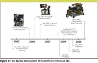
Figure 1
Initial work with the standoff instrument repeated studies performed in the laboratory at a standoff distance. We collected double-pulse LIBS spectra of RDX (cyclotrimethylenetrinitramine), Composition-B (36% TNT, 63% RDX, and 1% wax), Bacillus subtilis (BG), a mold sample (Alternia alternata), diisopropyl methyl phosphonate (C7H17O3P, DIMP, 96%), dimethyl methyl phosphonate (C3H9O3P, DMMP, 99%), diethyl ethyl phosphonate (C6H15O3P, DEEP, 98%), diethyl methyl phosphonate (C5H13O3P, DEMP, 98%), triethyl phosphate (C6H15O4P, TEP, 99%), oil, diesel fuel, road dust, house dust, fingerprint residue, and plastic (Type V polypropylene [C3H6]n). We also confirmed that double-pulse LIBS at a standoff distance was advantageous for explosives discrimination compared to single-pulse LIBS (36,44). To discriminate between a hazardous material and a benign substance, we have employed a variety of discrimination techniques ranging from simple linear correlation to more advanced chemometric techniques (43–45). So far, we have found that partial least squares discriminant analysis (PLS-DA) is the most effective chemometric method for classifying the hazardous residue samples. Expanding upon these initial studies, we collected standoff LIBS spectra of another large set of bioagent simulants, interferents, and chemical warfare agent simulants (37). We used PLS-DA to achieve discrimination between bioagent simulants and interferents as well as the five nerve agent simulants. In addition, a combined PLS-DA model was developed that incorporated all three classes of hazardous threats — chemical, biological, and explosive (CBE). In a limited study, the preliminary model yielded the following results: 100% true positives and a 2% false positive detection rate (37).
Standoff Systems at ARL
Prototype ST-LIBS systems were developed by ARL in conjunction with partners Ocean Optics, Inc. (Dunedin, Florida) and Applied Photonics Ltd. (Skipton, North Yorkshire, U.K.). The timeline for the development of these systems is shown in Figure 1. With each generation, significant design improvements have been made. The following section describes the design and capabilities of the standoff LIBS systems.
The first-generation system (Gen 1) incorporates a double-pulse Nd:YAG laser system (Big Sky CFR400-PIV, 1064 nm, 2 Hz, 250 mJ/ pulse, <10 ns pulse width). The lasers were chosen for their small footprint and rugged design. A commercially available 8" Schmidt–Cassegrain telescope (Meade LX200GPS) is used to collect the LIBS emission along the same path traversed by the laser ablation beam. The combined double-laser pulse is directed along the axis of the telescope by an articulating arm, enabling a full range of motion on the telescope for ease in targeting the sample. A diode laser (632 nm) illuminates the target spot coincident with the main IR laser. The infrared double-pulse beam is expanded with a simple two-lens system and is focused down range by a 3" positive lens (f = 475 mm). Plasma light collected by the telescope is focused into a fiber optic and sent to a gated CCD spectrometer (500–900 nm) developed by Ocean Optics. A digital camera and wireless range finder enable remote viewing and measurement of the distance to the target.
Although we were able to collect spectra of metals and explosive residues on metals at 20 m with the Gen 1 system, we found that the less-than-ideal beam quality of the lasers in the far-field resulted in a weaker plasma that made obtaining LIBS spectra of organic materials extremely challenging. For the Gen 2 system, therefore, a double-pulse laser source (Quantel Brilliant Twins, 1064 nm, 10 Hz, 335 mJ/ pulse, 5 ns pulse width) that provided superior beam quality (M2 < 2) and power at 20+ m was selected. As with the Gen 1 system, the two laser beams are combined before entering the articulating arm. A 14" telescope (Meade LX200GPS) was fitted with UV-coated optics to provide greater light-gathering power compared with Gen 1 and full broadband (UV–vis–NIR) capability. A custom-made three-channel gated CCD spectrometer (Ocean Optics) provides high light throughput and sensitivity from 190 nm to 840 nm. The entire system is mounted on a wheeled cart and is easily transportable. The data presented in the following section were acquired using the Gen 2 system (shown in Figure 1). The optimal timing parameters for the system are as follows: delay time tdelay = 2 μs, integration time gate tint = 100 μs, and interpulse separation Δt = 3 μs.
A third-generation standoff LIBS system has been designed to address the issue of eye safety. A single Nd:YAG laser (Quantel Brilliant B, 1064 nm, 10 Hz, 850 mJ, 6 ns pulse width) is shifted to 1.54 μm using a CH4/Ar-filled Raman cell developed by the National Center for Atmospheric Research (NCAR). As with the Gen 2 system, a modified 14" telescope is used to collect the light emitted from the laser-induced plasma. Testing of this system is under way, but this system will be used primarily as a proof-of-concept due to power limitations at 1.54 μm. The high laser energies necessary for these standoff systems will always be unsafe in the beam path, regardless of wavelength. However, potential damage from reflections and scatter can be mitigated by using wavelengths such as 1.54 μm or the third and fourth harmonics of the Nd:YAG rather than the fundamental and second harmonic of the Nd:YAG. Therefore, the area around the standoff system that must be controlled for laser safety is much smaller than if 1064 nm is used.
More recently, a fourth-generation standoff system has been designed and developed by Applied Photonics Ltd. and A3 Technologies LLC (Bel Air, Maryland) for recent field testing. A single Nd:YAG laser (Quantel Brilliant B, 1064 nm, 10 Hz, 850 mJ, 6 ns pulse width) and a modified 14" telescope are used to generate and collect light emission from the plasma. For this system, three Czerny–Turner spectrometers with ICCDs (Andor) are used to collect the LIBS spectra. Each spectrometer covers a portion of the LIBS spectrum. All of the standoff LIBS functions can be controlled from a remote desktop, including control of laser parameters, laser aiming, laser firing, spectrometer inputs, and data collection. In addition, several features have been automated such as autofocusing the laser, autofocusing the collection optics, and performing raster scans over a given area.
Finally, the newest generation (Gen5) standoff system developed by Applied Photonics, Ltd. was delivered to ARL in April 2009. This system is designed to work at 100+ m. It integrates all of the automation and computer control features of the Gen 4 system with two Nd:YAG lasers (Quantel Brilliant Bs, 1064 nm or 355 nm, 10 Hz, 850 mJ, 6 ns pulse width) for double-pulse operation. The Schmidt–Cassegrain telescope has been replaced by a military-spec 16" Ritchey–Chretien telescope (RC Optical Systems, Inc., Flagstaff, Arizona). A new broadband spectrometer (VSMS, PI-Acton, Trenton, New Jersey) with an ICCD will be used to collect the LIBS spectrum. The system will be evaluated using the same samples and discrimination methodology we have previously developed, but at much greater distances (>60 m).
Recent Work and Discussion
Our most recent work with explosives and bioagent surrogates has dealt with the influences background materials will have on the ability to discriminate residues. We have shown on simple aluminum substrates that standoff LIBS can successfully discriminate residues with similar elemental constituents. Explosive residue samples have been classified correctly 94% of the time (45). More complicated samples that contained simple mixtures of explosives and interferents were classified as explosives at an 80% true positive rate. Recently, we have investigated how using organic substrates rather than metallic substrates will affect our ability to discriminate between explosives and nonexplosive materials. The organic substrate material is ablated and entrained in the plasma; thus, the carbon and hydrogen contained in the organic substrate will interfere with the carbon and hydrogen atomic emission that originates from the explosive residue. We applied a small amount of RDX, road dust, and oil residue to a red silicone substrate to determine how well we could discriminate explosives from nonexplosives on an organic substrate. We collected 50 single-shot double-pulse LIBS spectra (at 25-m distance) of each residue on the substrate as well as the blank substrate itself. In Figure 2, a representative single-shot double-pulse spectrum of each residue on the organic substrate and the blank substrate are displayed. We used 40 of the 50 spectra for each residue and the blank to generate a model in PLS-DA. From each single-shot spectrum, nine intensities and 20 ratios of these intensities were calculated, as described in reference 44. These 29 variables were the inputs for the PLS-DA model. We calculated the model and then used the 10 remaining single-shot spectra from each residue type to test how the model performs. In Figure 3, which shows the predicted scores for the RDX class in the PLS-DA model for both the model and test spectra, all of the RDX residue on red silicone substrate test samples are above the classification threshold determined by the model and are thus classified as explosives. None of the nonexplosive residue test samples classify as explosive. These preliminary results indicate that LIBS can differentiate between explosive and nonexplosive residues on an organic substrate. Current work involves expanding the types of substrate materials and the number of explosive and interferent test samples to further test the PLS-DA models.
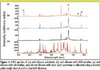
Figure 2
As described in the introduction, we have been investigating the use of LIBS with biological warfare agent surrogates for several years now. In fact, our first work with coupling chemometrics with LIBS data involved biomaterials (17). We also have performed an extensive study of biomaterials at a 20-m standoff range. These studies include an array of biological warfare agent surrogates, other biomaterials, and interferents. We demonstrated the successful discrimination of the anthrax surrogate BG (2% false negatives, 0% false positives) and ricin surrogate ovalbumin (0% false negatives, 1% false positives) (37). However, biological warfare agents can be grown in a variety of media. The elemental uptake of the bacterial spore will be different depending upon the growth media. Because LIBS tracks all of the elements in order to classify the material as a biological warfare agent or as an interferent, the growth media type could influence how the bioagent is classified. We obtained two spore types, BG and Bacillus thuringiensis (Bt), in two growth media, G-Media (G) and Sporulation Broth (SpBr) from ChemImage (Pittsburgh, Pennsylvania). We performed a small feasibility test on a limited sample set to observe the effect of growth media on the ability to discriminate between the two spores. We collected 10 single-shot LIBS spectra at 40 m for each spore type from each growth media and the blank aluminum substrate. In Figure 4, LIBS spectra of the biomaterials in the two growth media are displayed. In Figure 4b, there are several atomic emission lines that are only observed in the G growth media. In Figure 4c, there are observable differences in the relative intensities of the atomic emission lines for each spore type.
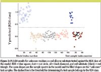
Figure 3
The goal was to be able to discriminate between Bt and BG irrespective of growth media. A number of atomic line intensities and ratios of the atomic emission intensities were selected as inputs into the PLS-DA model based upon our earlier work (37). Three classes were defined in the PLS-DA model — a BG class, a Bt class, and a substrate class (aluminum). Six BG in G samples and six BG in SpBr samples were used for the BG class; six Bt in G samples and six Bt in SpBr samples were used for the Bt class; and six aluminum substrates were used for the aluminum class in the model. The remaining 20 spectra were used as test samples for the PLS-DA model. In Figure 5, each combination of spore type and growth media test sample matches the correct spore class regardless of the growth media.
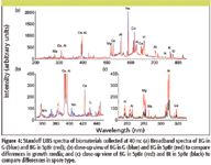
Figure 4
Conclusions
Despite substantial success in the laboratory and in the handful of field trials, standoff LIBS still needs further refinement before it can be considered a fully viable instrument for field use by military or civilian personnel. However, LIBS is one of only a handful of techniques that have been shown to detect explosive signatures at a standoff distance. Other techniques that are being investigated for standoff explosives detection include terahertz (THz) imaging, photofragmentation followed by laser-induced fluorescence (PF-LIF), photoacoustic spectroscopy, and Raman spectroscopy (46). THz time domain spectroscopy has been used to collect reflected absorption spectra of RDX at 30 m (47). Even at 30 m, however, atmospheric water vapor obstructs the RDX signal, and the interference will increase as standoff distance is increased (48,49). PF-LIF can be used as a standoff technique and has been demonstrated under laboratory conditions to detect TNT molecules at a distance of up to 2.5 m (50,51). A variation of photoacoustic spectrosocpy using quantum cascade lasers as the optical source for illuminating surface adsorbed explosives and quartz crystal tuning forks as the detectors has been demonstrated recently at a distance of 20 m (52). All of these techniques are still confined to the laboratory and are still in the early stages of development. While these other techniques have been applied to the detection of explosive materials at a standoff distance, to our knowledge, none have demonstrated discrimination between explosives and interferent materials at a standoff distance.
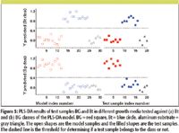
Figure 5
Raman spectroscopy is another optical technique that has demonstrated success for detecting explosive signatures (53). Raman can be used to obtain molecular information about the sample and does not leave an imprint, although photodegradation of the sample can occur. However, the signal intensity is weak and thus, acquisition time can be much longer relative to LIBS. Using LIBS and Raman together to provide orthogonal information is currently under investigation by several groups. The instruments conceivably can share the same laser and spectrometers while obtaining elemental and molecular information. The data can be fused from the various instruments to increase the probability of detection and decrease the false alarm rate.
Since 2004, standoff LIBS at ARL has progressed rapidly from simply obtaining spectra of residues to discriminating these materials from interferents under various conditions (such as distance, substrate, and laser parameters). Our recent work discriminating explosives on an organic substrate and biomaterials from different growth media has directly addressed potential challenges to standoff LIBS. Future work should address sample preparation as samples become more complex, model optimization for detection and discrimination of these samples, and the limits of detection (LOD) for a particular application. Increased complexity of samples presents a challenge for building and testing chemometric models. Consider a hypothetical 50/50 mixture of explosive and interferent distributed on a substrate. Numerous spectra of the mixture sample must be collected in order to populate the model. The distribution of explosive and interferent is not uniformly 50/50. Therefore, input into the model will contain spectral information from a wide range of explosive/interferent percentages. The same will hold true for test samples. In reference 45, the misclassification rate is much higher for mixtures. Some of this can be attributed to how the explosive mixtures were prepared and sampled. Some of the plasma interrogations that we classified as explosive might have sampled regions with little to no explosive. For a more valid test, the actual composition of each plasma interrogation must be known, both for the test sample and for the model samples. New ways of preparing samples for model building and testing must be developed. The model itself is also sensitive to changing experimental parameters. As seen in reference 45, changing the number of latent variables in a PLS-DA model will influence the number of false negatives and false positives. The number of classes and types of classes for a specific application can only be validated by testing with multiple iterations of test sets and adjustments to the contents of the model. As described previously, the model is only as good as the data that have been used to build the model. We must also determine the LOD. The LOD is highly dependent upon experimental parameters, including but not limited to target material, laser energy, focusing optics, collection efficiency, and substrate. It will also be dependent upon the ability to discriminate between the explosive and nonexplosive. Again, sample preparation is very important for determining the LOD. There must be a known quantity of the explosive that is consumed by the plasma. How these quantities are prepared as the concentration of the explosive decreases becomes an important question. Defining the LOD will be application-dependent as well as instrument-dependent.
Standoff LIBS, though still an evolving technology, is expected to have significant impact in real-world applications. The major attributes of LIBS, namely real-time response, no sample preparation required, and significant potential in sensitivity and specificity, are beginning to be applied at standoff distances of 100 m and beyond. In fact, due to the new generation of broadband, high-resolution spectrometers, standoff LIBS represents a new tool for general materials analysis, whether the material is hazardous or benign. Thus, the areas of application for standoff LIBS are much broader than just military and security. In the future, the performance of standoff LIBS will be improved due to advances in components, most notably in spectrometers and lasers. Such component improvements coupled with the increase in commercial capacity for building such systems suggests that in the next few years there will be many examples of real-world field applications.
Frank C. De Lucia, Jr., Jennifer L. Gottfried, Chase A. Munson, and Andrzej W. Miziolek are with the U.S. Army Research Laboratory, AMSRD-ARL-WM-BD, Aberdeen Proving Ground, Maryland.
The manufacturer of the software used in this research is Eigenvector, Wenatchee, Maryland.
References
(1) A. Miziolek, V. Palleschi, and I. Schechter, Eds., Laser Induced Breakdown Spectroscopy (Cambridge University Press, Cambridge, UK, 2006).
(2) D.A. Cremers and L.J. Radziemski, Handbook of Laser-Induced Breakdown Spectroscopy (John Wiley & Sons, Ltd., West Sussex, England, 2006).
(3) D.A. Rusak, B.C. Castle, B.W. Smith, and J.D. Winefordner, Crit. Rev. Anal. Chem. 27, 257–290 (1997).
(4) K. Song, Y.I. Lee, and J. Sneddon, Appl. Spectrosc. Rev. 32, 183–235 (1997).
(5) D.A. Rusak, B.C. Castle, B.W. Smith, and J.D. Winefordner, Trends Anal. Chem. 17, 453–461 (1998).
(6) J.D. Winefordner, I.B. Gornushkin, D. Pappas, O.I. Matveev, and B.W. Smith, J. Anal. At. Spectrom. 15, 1161–1189 (2000).
(7) J.D. Winefordner, I.B. Gornushkin, T. Correll, E. Gibb, B.W. Smith, and N. Omenetto, J. Anal. At. Spectrom. 19, 1061–1083 (2004).
(8) D.A. Cremers, Appl. Spectrosc. 41, 572–578 (1987).
(9) A. Ferrero and J.J. Laserna, Spectrochim. Acta, Part B 63, 305–311 (2008).
(10) B. Salle, P. Mauchien, and S. Maurice, Spectrochim. Acta, Part B 62B, 739–768 (2007).
(11) C. Lopez-Moreno, S. Palanco, and J.J. Laserna, J. Anal. At. Spectrom. 22, 84–87 (2007).
(12) S. Palanco, C. Lopez-Moreno, and J.J. Laserna, Spectrochim. Acta, Part B 61, 88–95 (2006).
(13) B. Sallé, P. Mauchien, J.-L. Lacour, S. Maurice, and R.C. Wiens, "Laser-Induced Breakdown Spectroscopy: A New Method for Stand-off Quantitative Analysis of Samples on Mars," in Lunar and Planetary Science XXXVI (Houston, Texas, 2005).
(14) B. Salle, J.-L. Lacour, E. Vors, P. Fichet, S. Maurice, D.A. Cremers, and R.C. Wiens, Spectrochim. Acta, Part B 59, 1413–1422 (2004).
(15) S. Palanco and J. Laserna, Rev. Sci. Instrum. 75, 2068–2074 (2004).
(16) F.C. DeLucia Jr., A.C. Samuels, R.S. Harmon, R.A. Walters, K.L. McNesby, A. LaPointe, R.J. Winkel Jr., and A.W. Miziolek, IEEE Sensors Journal 5, 681–689 (2005).
(17) A.C. Samuels, F.C. DeLucia Jr., K.L. McNesby, and A.W. Miziolek, Appl. Opt. 42, 6205–6209 (2003).
(18) F.C. DeLucia Jr., R.S. Harmon, K.L. McNesby, R.J. Winkel Jr., and A.W. Miziolek, Appl. Opt. 42, 6148–6152 (2003).
(19) C.A. Munson, J.L. Gottfried, E.G. Snyder, F.C. DeLucia Jr., B. Gullett, and A.W. Miziolek, Appl. Opt. 47, G48–G57 (2008).
(20) S. Rai, A.K. Rai, and S.N. Thakur, Appl. Phys. B 91, 645–650 (2008).
(21) Y. Dikmelik, C. McEnnis, and J.B. Spicer, Opt. Express 16, 5332–5337 (2008).
(22) J. Diedrich, S.J. Rehse, and S. Palchaudhuri, Appl. Phys. Lett. 90, 163901 (2007).
(23) J. Diedrich, S.J. Rehse, and S. Palchaudhuri, J. Appl. Phys. 102, 014702/014701–014702/014708 (2007).
(24) S.J. Rehse, J. Diedrich, and S. Palchaudhuri, Spectrochim. Acta, Part B 62, 1169–1176 (2007).
(25) J.D. Hybl, S.M. Tysk, S.R. Berry, and M.P. Jordan, Appl. Opt. 45, 8806–8814 (2006).
(26) J.D. Hybl, G.A. Lithgow, and S.G. Buckley, Appl. Spectrosc. 57, 1207–1215 (2003).
(27) M. Baudelet, J. Yu, M. Bossu, J. Jovelet, J.P. Wolf, T. Amodeo, E. Frejafon, and P. Laloi, Appl. Phys. Lett. 89, 163903 (2006).
(28) M. Baudelet, L. Guyon, J. Yu, J. P. Wolf, T. Amodeo, E. Frejafon, and P. Laloi, J. Appl. Phys. 99, 84701 (2006).
(29) M. Baudelet, L. Guyon, J. Yu, J. P. Wolf, T. Amodeo, E. Frejafon, and P. Laloi, Appl. Phys. Lett. 88, 063901 (2006).
(30) P. B. Dixon and D. W. Hahn, Anal. Chem. 77, 631–638 (2005).
(31) N. Leone, G. D'Arthur, P. Adam, and J. Amouroux, High Temp. Mater. Process. 8, 1–22 (2004).
(32) A.R. Boyain-Goitia, D.C.S. Beddows, B.C. Griffiths, and H.H. Telle, Appl. Opt. 42, 6119–6132 (2003).
(33) D.W. Merdes, J.M. Suhan, J.M. Keay, D.M. Hadka, and W.R. Bradley, Spectroscopy 22, 28–35 (2007).
(34) F.C. De Lucia Jr., J.L. Gottfried, C.A. Munson, and A.W. Miziolek, Spectrochim. Acta, Part B 62, 1399–1404 (2007).
(35) C.A. Munson, F.C. De Lucia Jr., T. Piehler, K.L. McNesby, and A.W. Miziolek, Spectrochim. Acta, Part B 60, 1217–1224 (2005).
(36) J.L. Gottfried, F.C. De Lucia Jr., C.A. Munson, and A.W. Miziolek, Spectrochim. Acta, Part B 62, 1405–1411 (2007).
(37) J.L. Gottfried, F.C. De Lucia Jr., C.A. Munson, and A.W. Miziolek, Appl. Spectrosc. 62, 353–363 (2008).
(38) E.G. Snyder, C.A. Munson, J.L. Gottfried, F.C. De Lucia Jr., B. Gullett, and A. Miziolek, Appl. Opt. 47, G80–G87 (2008).
(39) C. Gautier, P. Fichet, D. Menut, J.-L. Lacour, D. L'Hermite, and J. Dubessy, Spectrochim. Acta, Part B 60, 792–804 (2005).
(40) J. Scaffidi, S.M. Angel, and D.A. Cremers, Anal. Chem. 78, 24–32 (2006).
(41) M. Corsi, G. Cristoforetti, M. Giuffrida, M. Hidalgo, S. Legnaioli, V. Palleschi, A. Salvetti, E. Tognoni, and C. Vallebona, Spectrochim. Acta, Part B 59, 723–735 (2004).
(42) V.I. Babushok, F.C. De Lucia Jr., J.L. Gottfried, C.A. Munson, and A.W. Miziolek, Spectrochim. Acta, Part B 61, 999–1014 (2006).
(43) C. Lopez-Moreno, S. Palanco, J. Javier Laserna, F. De Lucia, Jr., A.W. Miziolek, J. Rose, R.A. Walters, and A.I. Whitehouse, J. Anal. At. Spectrom. 21, 55–60 (2006).
(44) J.L. Gottfried, F.C. De Lucia Jr., C.A. Munson, and A.W. Miziolek, J. Anal. At. Spectrom. 23, 205–216 (2008).
(45) F.C. De Lucia Jr., J.L. Gottfried, C.A. Munson, and A.W. Miziolek, Appl. Opt. 47, G112–G121 (2008).
(46) C.A. Munson, J.L. Gottfried, F.C. De Lucia Jr., K.L. McNesby, and A.W. Miziolek, "Laser-based Detection Methods of Explosives," in Counterterrorist Detection Techniques of Explosives, J. Yinon, Ed. (Elsevier, Amsterdam, 2007), pp. 279–321.
(47) H. Zhong, Terahertz Wave Reflective Sensing and Imaging (Rensselaer Polytechnic Institute, Troy, New York, 2006).
(48) M. van Exter, C. Fattinger, and D. Grischkowsky, Opt. Lett. 14, 1128-1130 (1989).
(49) T. Yuan, H.B. Liu, J.Z. Xu, F. Al-Douseri, Y. Hu, and X.-C. Zhang, Proc. SPIE 5070, 28–37 (2003).
(50) T. Arusi-Parpar, D. Heflinger, and R. Lavi, Appl. Opt. 40, 6677–6681 (2001).
(51) D. Heflinger, T. Arusi-Parpar, Y. Ron, and R. Lavi, Opt. Comm. 204, 327–331 (2002).
(52) C.W. Van Neste, L.R. Senesac, and T. Thundat, Appl. Phys. Lett. 92, 234102–234103 (2008).
(53) J.C. Carter, S.M. Angel, M. Lawrence-Snyder, J. Scaffidi, R.E. Whipple, and J.G. Reynolds, Appl. Spectrosc. 59, 769–775 (2005).

LIBS Illuminates the Hidden Health Risks of Indoor Welding and Soldering
April 23rd 2025A new dual-spectroscopy approach reveals real-time pollution threats in indoor workspaces. Chinese researchers have pioneered the use of laser-induced breakdown spectroscopy (LIBS) and aerosol mass spectrometry to uncover and monitor harmful heavy metal and dust emissions from soldering and welding in real-time. These complementary tools offer a fast, accurate means to evaluate air quality threats in industrial and indoor environments—where people spend most of their time.
NIR Spectroscopy Explored as Sustainable Approach to Detecting Bovine Mastitis
April 23rd 2025A new study published in Applied Food Research demonstrates that near-infrared spectroscopy (NIRS) can effectively detect subclinical bovine mastitis in milk, offering a fast, non-invasive method to guide targeted antibiotic treatment and support sustainable dairy practices.
Smarter Sensors, Cleaner Earth Using AI and IoT for Pollution Monitoring
April 22nd 2025A global research team has detailed how smart sensors, artificial intelligence (AI), machine learning, and Internet of Things (IoT) technologies are transforming the detection and management of environmental pollutants. Their comprehensive review highlights how spectroscopy and sensor networks are now key tools in real-time pollution tracking.