Topographic and 3D Raman Imaging
Application Notebook
Confocal Raman imaging opened the door for many applications in Raman spectroscopy and imaging that were previously unavailable for measurement with conventional (non-confocal) Raman methods.
Confocal Raman imaging opened the door for many applications in Raman spectroscopy and imaging that were previously unavailable for measurement with conventional (non-confocal) Raman methods. However, high confocality always results in a high focus sensitivity and this can make measurements difficult with rough or inclined samples. The TrueSurface imaging extension resolves these problems and extends Raman imaging to large scale (> 1×1 mm2) samples without extensive tilt alignment or sample preparation and without comprising the advantages of confocal imaging. For the characterization of the inner properties of a sample with Raman Spectroscopy an ultrasensitive confocal Raman microscope allows the acquisition of a Raman image stack revealing 3D information on the distribution of the chemical compounds.
TrueSurface Microscopy for Topographic Raman Imaging
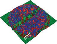
WITec's exclusive True Surface Microscopy mode makes it possible to perform confocal imaging measurements parallel with and guided by large area topographic scans (> 1×1 mm2). To achieve this unique capability, the WITec microscope series can be equipped with a highly precise sensor for optical profilometry. The large area topographic coordinates from the profilometer measurement are used to perfectly trace the samples surface in either confocal or confocal Raman imaging mode. Sample preparation is reduced to a minimum without having to compromise the confocality of the system. The TrueSurface Microscopy mode is beneficial for many applications, including the characterization of micromechanical, medical, or semiconductor devices, the mapping of functionalized surfaces, or the imaging of bio-medical or pharmaceutical material surface properties. Figure 1 shows a height profile of a pharmaceutical tablet (aspirin) with the confocal Raman measurement overlaid. The drugs are labeled red and blue respectively, while the excipient is shown in green. Topographic variation was more than 300 µm.

Figure 1: Topographic Raman image of an aspirin tablet obtained with the TrueSurface Microscopy Mode.
Confocal 3D Raman Imaging
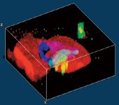
By integrating a Raman spectrometer within a state-of-the-art confocal microscope setup, Raman imaging with a spatial resolution down to 200 nm laterally and 500 nm vertically can be achieved using visible light excitation. The sample is scanned point-by-point and line-by-line, and at every image pixel a complete Raman spectrum is taken while continuously scanning. These multi-spectrum files are then analyzed to display the distribution of chemical sample properties. By taking a stack of images with different focal positions, the geometry of samples can be reconstructed in 3D. Figure 2 shows such a 3D Raman Image of a fluid inclusion in garnet (60×60×30 µm) with the fluid inclusion (water) displayed in blue and the garnet in red. Typical integration times in a confocal Raman microscope are between 700 µs and 100 ms per spectrum, so that a complete Raman image consisting of tens of thousands of spectra takes between a few seconds and 20 min.

Figure 2: 3D Confocal Raman image of a fluid inclusion in garnet (60Ã60Ã30 µm; red: garnet, blue: water, green: calcite, turquoise: mica).
WITec GmbH
Lise-Meitner-Str. 6, 89091 Ulm, Germany
tel: +49 (0) 731 140 700, E-mail: info@witec.de
Website: www.witec.de

Thermo Fisher Scientists Highlight the Latest Advances in Process Monitoring with Raman Spectroscopy
April 1st 2025In this exclusive Spectroscopy interview, John Richmond and Tom Dearing of Thermo Fisher Scientific discuss the company’s Raman technology and the latest trends for process monitoring across various applications.
A Seamless Trace Elemental Analysis Prescription for Quality Pharmaceuticals
March 31st 2025Quality assurance and quality control (QA/QC) are essential in pharmaceutical manufacturing to ensure compliance with standards like United States Pharmacopoeia <232> and ICH Q3D, as well as FDA regulations. Reliable and user-friendly testing solutions help QA/QC labs deliver precise trace elemental analyses while meeting throughput demands and data security requirements.