Exploring Neurochemistry Using Raman Spectroscopy
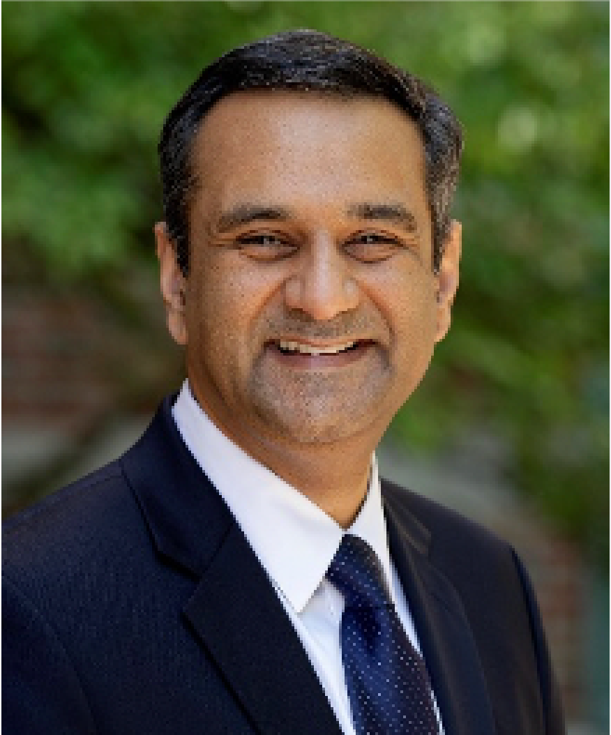
Bhavya Sharma is an assistant professor of chemistry at the University of Tennessee in Knoxville, where she is conducting research to detect active and important biomolecules related to hormone regulation, neurological health, and disease diagnosis. For this work, she is applying various forms of Raman spectroscopy, including surface-enhanced Raman spectroscopy (SERS) spatially offset Raman spectroscopy (SORS), and surface-enhanced spatially offset Raman spectroscopy (SESORS). She is the winner of the 2021 Emerging Leader in Molecular Spectroscopy Award, which is presented by Spectroscopy magazine. This annual award recognizes the achievements and aspirations of a talented young molecular spectroscopist, selected by an independent scientific committee. We recently interviewed her about her work.
As a postdoctoral researcher, you were able to combine SERS and SORS into a hybrid analytical technique referred to as SESORS (1). Would you explain to our readers how this concept came about, and what is the ultimate purpose of this technique?
I was not actually the first person to combine SERS and SORS. This was done by Pavel Matousek, Nick Stone, Karen Faulds, and Duncan Graham in the United Kingdom. Karen is the person who came up with the acronym SESORS. My contribution to innovative applications of SESORS was to be the first person to apply it towards Raman detection through bone. When President Obama announced the Brain Initiative in 2013, I started thinking about how we could apply Raman spectroscopy to looking at the brain. I used to have discussions with my postdoctoral advisor about this, but it seemed that everything would require removing pieces of the skull or drilling holes into the skull to take Raman measurements. I was familiar with SORS, which had been used to measure signals through skin and tissue, and I thought, why not through bone? I wasn’t actually sure it would work. Bone is dense; it is more difficult to get light through it, and more importantly, to get the Raman scattered light back out. I decided to start trying to take measurements, and I didn’t tell my postdoctoral advisor about it. I wanted to see if I could make it work before I told him about it. My first test was actually to use eggs, since an eggshell has a similar chemical composition as bone. I thought if I could detect a SERS signal through the eggshell, then maybe I could do it through bone, and it worked! I moved onto to bone after that.
The ultimate purpose of this technique is to develop a way to measure neurochemicals related to neurological disease and brain injury in the brain without having to drill holes in or remove pieces of the skull. Many neurological conditions and diseases are not diagnosed until a person starts displaying symptoms. By that point, it may be too late to treat the actual disease because of degeneration of neurons, which do not grow back. We are working to develop methods for noninvasive detection of early onset neurological disease.
By combining Raman techniques and chemometrics, you were able to directly detect important biomolecules, such as cortisol, for the first time (2). Why is this research important? What were the major analytical hurdles you encountered when developing a rapid non-invasive method to detect cortisol at physiological concentrations?
After the last year and a half, everyone knows what high stress levels feel like. We are working to develop methods for detecting cortisol in noninvasively collected biofluids, particularly to help monitor cortisol in people with high stress jobs, like first responders, military personnel, and so forth. We want something that can be used in the field, so that a person can be evaluated in the middle of a high stress situation, and a decision can be made if they need some time away. High levels of cortisol in your system for extended periods of time can have long-term negative health consequences.
The major hurdle we encountered is that cortisol is not very soluble! The body is amazing. Somehow, we have cortisol in our systems, which are primarily made of water, salts, and proteins. It took us a while to figure out how to get cortisol into physiologically relevant solvents.
Some of your most influential and instructive research publications may very well be in your recent review articles on the explanations and applications of Raman phenomena (3–5). What are the main takeaways you have learned from researching and writing these review articles?
For me, it is just so great to see such vast applications of Raman spectroscopy. We have so many great researchers around the world developing new substrates for SERS, to help increase enhancement, as well as new Raman spectroscopy-based methods for solving a range of problems from biological, environmental, and energy, to name a few. It is really exciting to read about and highlight all of the Raman spectroscopy-related research efforts.
You have also published recent work measuring volatile organic compound (VOC) detection using a porous-silicon-oxide coated disc-on-pillar array (6). Can you explain the significance and meaning of this work?
We collaborated with researchers at Oak Ridge National Laboratory to utilize these disc-on-pillar arrays for SERS gas sensing. We were interested in developing a SERS-based method for detecting VOCs in two ways, one for industrial applications where you could monitor VOCs at low concentrations; the other, which was somewhat of a crazy idea, was for human health monitoring. It turns out that, depending on your health, you can exhale something like a thousand different chemicals on your breath, some of which are VOCs. The idea was to create a SERS-based sensor for some of these human health-related VOCs. The work you mention here was a first step towards developing these sensors. This is something we’re still working on developing.
What were some of the key challenges you have encountered during your work? What would you consider to be the most useful contributions of your work?
The main experimental challenges we’ve faced have centered around the idea of how do we detect biological molecules of interest at the concentration ranges that are physiologically relevant. Resolving these challenges is where SERS comes in, because it allows us to get to the low concentration ranges. However, we’ve run into issues with SERS where some molecules will not get close enough to the surface to be enhanced, so then we need to come up with ways to address that. Luckily, we have the literature to find ways to address these issues, including things like using antibodies or other capture molecules to help bring the analyte molecules within the sensing range of the SERS substrates. Other issues challenges have included things like how to detect signals through thicker areas of bone. The human skull is between 3–14 mm thick depending on where you measure on the skull, so we’ve demonstrated SESORS detection through almost 3 mm of bone. How do we push that detection through thicker areas? This question is one thing we’re currently working on answering.
I think the most useful contributions of our work include showing that you can use SESORS to measure Raman scattering through bone. Other groups have followed us in this, particularly for detection of glioblastomas, an aggressive type of cancer, in the brain. We also published an extensive study of SERS detection of neurotransmitters on two different metals (gold and silver) at three different excitation wavelengths, which we thought would be helpful for researchers in the field since, when we first started looking at neurotransmitters, the literature contained either older studies or conflicting results.
What are some key aspects in science that motivate you? Would you share some of your work and organizational habits that have helped you be productive and successful professionally?
I’m really motivated by the problem-solving aspect of science. There are so many issues in this world that need creative solutions. I am driven by being part of the solution. I also am a huge proponent of Raman spectroscopy. I sometimes think it can be used to solve anything; it really can’t, but I definitely like to try to solve various problems applying different forms of Raman spectroscopy. Another thing that motivates me is helping students learn. My group always has a large number of undergraduate researchers contributing to our work. I encourage undergraduate students to pursue research, and if I don’t have space in my group, I try to help them find research positions in other groups. I think being part of a research group is an invaluable experience for an undergraduate student.
One of the first things I learned as a new assistant professor was to turn the notifications from my email off. I try to set aside specific times to look at email, otherwise it can be overwhelming and really distracting. My group also implemented the use of Slack before the pandemic. I had tried to use it early in my career when I had two graduate students, and it didn’t really work well. Now, however, with five graduate and eight undergraduate students, it has provided us with a great tool for communicating, particularly while we were all working from home. We’ve also implemented different project management tools to help people keep track of timelines. Another thing I learned personally during the pandemic that has really helped me be more productive is that I need to set aside time to just think, which sounds weird to say, but it is really easy to get caught up in emails, teaching, talking to students about research, and so on. But to come up with new ideas or to solve problems we’re having; I need quiet time to read literature or just think about things.
What can you share with our readers regarding your next area of interest for your research?
We’re still working on improving methods for non-invasive detection of neurochemicals in the brain. This will be something we’re working on for the foreseeable future. We also are always looking for new ways to combine analytical techniques with Raman spectroscopy to help improve things like limits of detection.
Do you have any words of advice for young people desiring a career in science?
Find what makes you really excited and happy. Try out different things to figure that out. Get involved in undergraduate research; if you’re still in high school or just starting college, don’t think you need to wait until your senior year to do this. You can start even as a freshman.
References
(1) A.S. Moody, P.C. Baghernejad, K. Webb, and B. Sharma, Anal. Chem. 89(11), 5688–5692 (2017).
(2) T.J. Moore, and B. Sharma, Anal. Chem. 92(2), 2052–2057 (2019).
(3) T.J. Moore, A.S. Moody, T.D. Payne, G.M. Sarabia, A.R. Daniel, and B. Sharma, Biosensors 8(2), 46 (2018).
(4) B. Sharma, R.R. Frontiera, A.I. Henry, E. Ringe, and R.P. Van Duyne, Mater. Today 15(1-2), 16–25 (2012).
(5) M.D. Sonntag, J.M. Klingsporn, A.B. Zrimsek, B. Sharma, L.K. Ruvuna, and R.P. Van Duyne, Chem. Soc. Rev. 43(4), 1230–1247 (2014).
(6) T.J. Moore, and B. Sharma, “Volatile Organic Compound Detection using Porous-Silicon-Oxide Coated Disc-on-Pillar Arrays, In Optical Sensors” (pp. SeTu4E-4). Optical Society of America (July, 2018). https://doi.org/10.1364/SENSORS.2018.SeTu4E.4
Bhavya Sharma is an assistant professor of chemistry at the University of Tennessee in Knoxville. She has developed novel Raman spectroscopy methods for neurochemical detection, including surface-enhanced spatially offset Raman spectroscopy (SESORS). Her group has demonstrated detection of neurochemicals through the skull, down to nanomolar concentration ranges. Additionally, she is developing methods to demonstrate direct detection of molecules, such as cortisol, for the first time by combining SERS and multivariate analysis. ●


How Do We Improve Elemental Impurity Analysis in Pharmaceutical Quality Control?
May 16th 2025In this final part of our conversation with Harrington and Seibert, they discuss the main challenges that they encountered in their study and how we can improve elemental impurity analysis in pharmaceutical quality control.
How do Pharmaceutical Laboratories Approach Elemental Impurity Analysis?
May 14th 2025Spectroscopy sat down with James Harrington of Research Triangle Institute (RTI International) in Research Triangle Park, North Carolina, who was the lead author of this study, as well as coauthor Donna Seibert of Kalamazoo, Michigan. In Part I of our conversation with Harrington and Seibert, they discuss the impact of ICH Q3D and United States Pharmacopeia (USP) <232>/<233> guidelines on elemental impurity analysis and how they designed their study.
Nanometer-Scale Studies Using Tip Enhanced Raman Spectroscopy
February 8th 2013Volker Deckert, the winner of the 2013 Charles Mann Award, is advancing the use of tip enhanced Raman spectroscopy (TERS) to push the lateral resolution of vibrational spectroscopy well below the Abbe limit, to achieve single-molecule sensitivity. Because the tip can be moved with sub-nanometer precision, structural information with unmatched spatial resolution can be achieved without the need of specific labels.
The Rise of Smart Skin Using AI-Powered SERS Wearable Sensors for Real-Time Health Monitoring
May 5th 2025A new comprehensive review explores how wearable plasmonic sensors using surface-enhanced Raman spectroscopy (SERS) are changing the landscape for non-invasive health monitoring. By combining nanotechnology, AI, and real-time spectroscopy analysis to detect critical biomarkers in human sweat, this integration of nanomaterials, flexible electronics, and AI is changing how we monitor health and disease in real-time.

.png&w=3840&q=75)

.png&w=3840&q=75)


.png&w=3840&q=75)


.png&w=3840&q=75)


