Infrared Spectroscopy of Polymers XII: Polyaramids and Slip Agents
After introducing organic nitrogen polymers in the last column, we continue this theme by studying an important family of polymers—polyaramids. In this column, we examine the spectra of polyaramids, which are polyamides that contain an aromatic ring in their chain. Included in this family is Kevlar, which is an important polymer many are familiar with. We also examine the spectra of “slip agents,” which are small molecule amides used to lubricate injection molds and are oftentimes found as contaminants in polymer products.
A common way of forming polymers into useful products is called injection molding (1). In this process, melted polymer is injected into a cavity or mold in the shape of a desired product. The melted plastic conforms to the shape of the mold; once the plastic cools, the mold is opened and the newly made product is ejected. The process is then repeated. Injection molding is a fast and inexpensive way of making many plastic items (1).
However, a problem with injection molding is that the manufactured product can sometimes stick to the surface of the mold, meaning the process must be shut down and the stuck piece removed, which wastes time and money. A trick injection molders use to solve this problem is to coat the inside of the mold with a slip agent, a material that lubricates the surface of the mold, allowing formed plastic products to be ejected from the mold more easily.
Small molecule amides are frequently used as slip agents. The reason I am including these in a discussion of polymers is that slip agents can be found as surface contaminants on some types of polymers, and their spectra can interfere with determining the structure of the polymer itself. For example, a polyethylene-based product with an amide slip agent on its surface will exhibit peaks from the polymer and the amide, adding confusion as to whether the product is made from a polyamide, polyethylene, or both. This confusion is a problem with surface sensitive infrared (IR) sampling techniques, such as attenuated total reflectance (ATR), where the slip agent on the surface is more readily sampled than the underlying polymer because of the small depth of penetration of the technique (2). A way around this problem is to simply clean the surface of the polymer with a paper towel soaked in solvent before taking the spectrum of the sample.
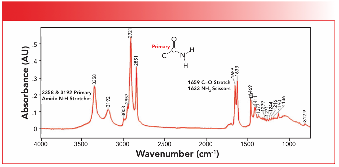
The mid-IR spectrum of a slip agent containing a primary amide group, and the structural framework of the primary amide group, are seen in Figure 1.
FIGURE 1: The IR spectrum of a slip agent containing a primary amide group. The structural framework of the primary amide group is also shown.
Remember from our last article that amides come in three varieties: primary; secondary; and tertiary (3). The quantity being counted is the number of C-N bonds, and we saw that a primary amide has one C-N bond, a secondary amide has two C-N bonds, and a tertiary amide has three C-N bonds (3). Additionally, as the number of C-N bonds goes up, the number of N-H bonds goes down. Thus, a primary amide contains an NH2 group, a secondary amide contains a N-H group, and tertiary amides have no N-H bonds. We have also seen that most polyamides contain secondary amide linkages in their backbones (3).
We have discussed the IR spectra of primary amides previously (4). The group wavenumbers for primary amides are shown in Table I.
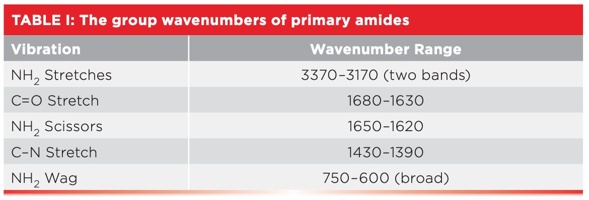
Briefly, primary amides exhibit two N-H stretching peaks between 3370 and 3170 cm-1 (going forward, all peak positions will be in cm-1 units even if not so noted), a carbonyl stretch from 1680 to 1630 as exhibited by all amides, a NH2 scissors peak from 1650 to 1620, and a N-H wag from 750 to 600 (4).
We know the slip agent whose spectrum appears in Figure 1 is a primary amide, because of the two N-H stretches at 3358 and 3192, and the C=O stretch at 1659. These three peaks comprise the diagnostic group wavenumber pattern for primary amides. Note that the N-H stretches in Figure 1 fall in the same range as O-H stretches, but are narrower and less intense than the O-H stretches we are used to seeing. This is because hydrogen bonding for N-H bonds is weaker than for O-H bonds, ultimately because of the fact that O-H bonds are more polar than N-H bonds (5). The NH2 scissors peak in Figure 1 is seen at 1633. There should be a primary amide NH2 wagging peak in Figure 1. However, the spectrum in Figure 1 was measured using an ATR crystal that cut off at 750—thus, we cannot see this peak.
Polyaramids
The word aramid is short for aromatic amide (6). A polyaramid, then, is a polymer with an aromatic amide in its backbone. If the polymer chain contains a para linkage, it is called a para-aramid, whereas, if the chain contains a meta linkage, it is called a meta-aramid (6).
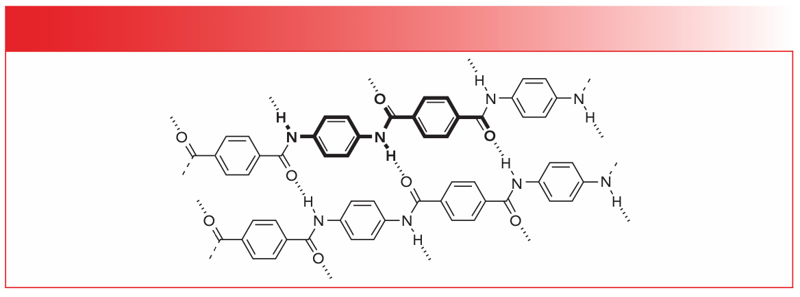
A well-known para-polyaramid is Kevlar, which is a tough, light, and strong polymer made into bulletproof vests and body armor, among other things. The key to the strength of aramids is that adjacent polymer chains can hydrogen bond through the N-H and C=O functional groups, as illustrated in Figure 2.
FIGURE 2: The hydrogen bonding that takes place in the para-polyaramid Kevlar, which is responsible for its high strength.
A single hydrogen bond may not be very strong, but moles upon moles of them are, which gives materials like Kevlar their strength. Similarly, cellulose chains interact via OH/OH hydrogen bonds, strengthening their fibers and allowing such magnificent structures as trees to rise hundreds of feet into the air (7).
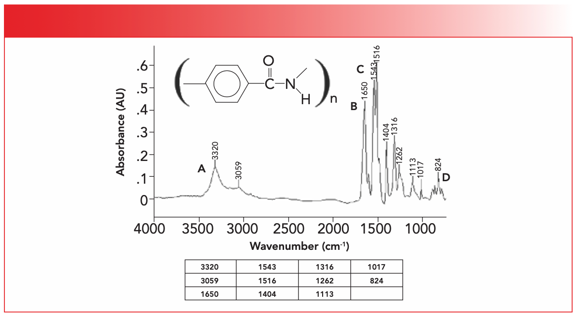
The IR spectrum of Kevlar, whose proper chemical name is polyparaphenylene terephthalamide, is seen in Figure 3.
FIGURE 3: The IR spectrum of a fiber of the polyaramid Kevlar, measured using an IR microscope. The proper chemical name for this material is polyparaphenylene terephthalamide.
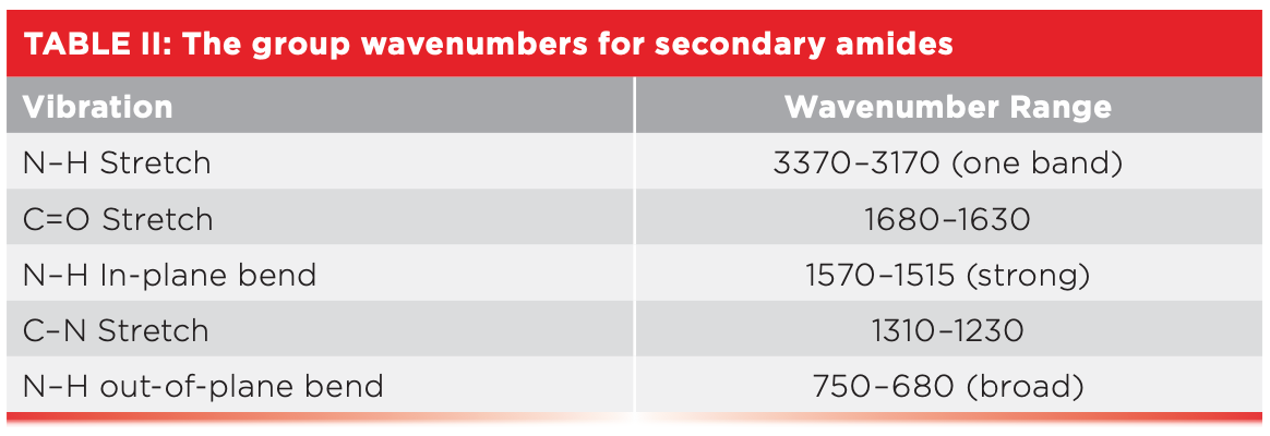
Similar to most polyamides, the Kevlar backbone contains secondary amide linkages. Recall from last time that diagnostic group wavenumber pattern of peaks for secondary amides are one N-H stretching peak from 3370 to 3170, a C=O stretching peak from 1680 to 1630, and an unusually intense N-H in-plane bending peak from 1570 to 1515 (3). Table II shows the group wavenumbers for secondary amides.
In Figure 3, there is a single N-H stretching peak labeled A at 3320, as expected for a secondary amide (3). Note that this peak is of medium width and intensity, and it is much smaller and narrower than a typical O-H stretching peak. The C=O stretch for Kevlar is labeled B at 1650, in the correct carbonyl stretching range for all amides. I assign the neighboring peak labeled C at 1543 as the N-H in-plane bending peak. Now, there is also an intense peak at 1516 that could also be assigned as the N-H in-plane bend. I assigned the peak at 1543 as the N-H in-plane bend based on experience; it has been my observation for a large variety of polyamides, including nylons, that the N-H in-plane bend falls approximately at 1540. We saw this last time in the spectrum of Nylon-6,6 (3). My assignment of the 1516 peak is a benzene ring “ring mode” (8). The broadened envelope labeled D in Figure 3 is the N-H out-of-plane bend or wag. The sharp peak on top of this envelope at 824 is a para-ring out-of-plane C-H bending vibration (9).
The IR spectrum and structure of a meta-polyaramid, Nomex, is seen in Figure 4. Nomex is used to make fireproof suits, among other things (10). Note that in the structure in Figure 4, there is one hydrogen separating the bonds attached to the benzene ring, making this a metagroup (9).
FIGURE 4: The IR spectrum and chemical structure of the meta-polyaramid Nomex.
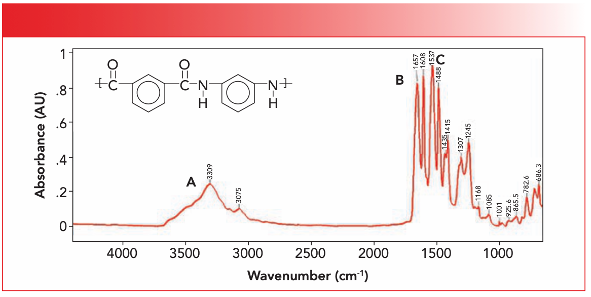
Like all the other polyamides whose spectra we have examined, Nomex contains a secondary amide linkage in its backbone. Consulting Table II, we find that the expected single N-H stretch labeled A is at 3309, the C=O stretch labeled B is at 1657, and the N-H in-plane is at 1537, sticking to my idea that many that many polyamides exhibit their N-H in plane bend around 1540. The N-H wagging envelope is seen as a rise at the end of the spectrum, with some sharp peaks on top of it that we will discuss below.
The intense peak at 1608 is noteworthy, and I assign it as a benzene ring mode vibration, similar to the peak at 1516 in Figure 3. Recall that metasubstituted benzene rings have a ring mode peak at 690±10, and a C-H wagging peak from 810 to 750 (9). In Figure 4, the ring mode is at 686 and the C-H wag is at 782. Both of these sharp peaks are riding on top of the broader N-H wagging envelope.
Conclusions
Slip agents are commonly used amide small molecules that lubricate the molds in injection molding processes. We studied the spectra of one of these, a primary amide, and discussed how they can contaminate the spectra of polymeric materials. Polyaramids are polyamides with aromatic rings in their backbones. We studied the spectrum of a para-polyamide, the well-known polymer Kevlar, and of a meta-polyaramid, Nomex. All these materials follow the group wavenumber rules for primary and secondary amides we have discussed previously.
References
(1) Wikipedia. Injection moulding. https://en.wikipedia.org/wiki/Injection_moulding (accessed 2023-04-18)
(2) Smith, B. C. Fundamentals of Fourier Transform Infrared Spectroscopy, 2nd Ed., CRC Press, Boca Raton, 2011.
(3) Smith, B. C. Infrared Spectroscopy of Polymers, XI: Introduction to Organic Nitrogen Polymers. Spectroscopy 2023, 38 (3), 14–18. DOI: 10.56530/spectroscopy.vd3180b5
(4) Smith, B. C. Organic Nitrogen Compounds, VII: Amides—The Rest of the Story. Spectroscopy 2020, 35 (1), 10–15.
(5) Smith, B. C. Organic Nitrogen Compounds, Part I: Introduction. Spectroscopy 2020, 34 (1), 10-15.
(6) Wikipedia. Aramid. https://en.wikipedia.org/wiki/Aramid (accessed 2023-04-18)
(7) Smith, B. C. The Infrared Spectra of Polymers, VI: Polymers With C-O Bonds. Spectroscopy 2022, 37 (5), 15-19,27. DOI: 10.56530/spectroscopy.ly3071f5
(8) Smith, B. C. Group Wavenumbers and an Introduction to the Spectroscopy of Benzene Rings. Spectroscopy 2016, 31 (3), 34-37.
(9) Smith, B. C. Distinguishing Structural Isomers: Mono- and Disubstituted Benzene Rings. Spectroscopy 2016, 31 (5), 36-39.
(10) Wikipedia. Nomex. https://en.wikipedia.org/wiki/Nomex (accessed 2023-04-18)
Brian C. Smith, PhD, is the founder and CEO of Big Sur Scientific, a maker of portable mid-infrared cannabis analyzers. He has over 30 years experience as an industrial infrared spectroscopist, has published numerous peer-reviewed papers, and has written three books on spectroscopy. As a trainer, he has helped thousands of people around the world improve their infrared analyses. In addition to writing for Spectroscopy, Dr. Smith writes a regular column for its sister publication Cannabis Science and Technology and sits on its editorial board. He earned his PhD in physical chemistry from Dartmouth College. He can be reached at: SpectroscopyEdit@MMHGroup.com ●

AI Shakes Up Spectroscopy as New Tools Reveal the Secret Life of Molecules
April 14th 2025A leading-edge review led by researchers at Oak Ridge National Laboratory and MIT explores how artificial intelligence is revolutionizing the study of molecular vibrations and phonon dynamics. From infrared and Raman spectroscopy to neutron and X-ray scattering, AI is transforming how scientists interpret vibrational spectra and predict material behaviors.
Real-Time Battery Health Tracking Using Fiber-Optic Sensors
April 9th 2025A new study by researchers from Palo Alto Research Center (PARC, a Xerox Company) and LG Chem Power presents a novel method for real-time battery monitoring using embedded fiber-optic sensors. This approach enhances state-of-charge (SOC) and state-of-health (SOH) estimations, potentially improving the efficiency and lifespan of lithium-ion batteries in electric vehicles (xEVs).
New Study Provides Insights into Chiral Smectic Phases
March 31st 2025Researchers from the Institute of Nuclear Physics Polish Academy of Sciences have unveiled new insights into the molecular arrangement of the 7HH6 compound’s smectic phases using X-ray diffraction (XRD) and infrared (IR) spectroscopy.