High-Definition Fourier Transform Infrared Spectroscopic Imaging of the Tumor Microenvironment
In cancer, the tissue microenvironment is an important determinant of disease progression and development. Spectroscopic imaging offers promise to reveal important information about what is happening in these microenvironments, with the potential to better predict tumor behavior. Professor Rohit Bhargava and his team at the University of Illinois, where they have established the Cancer Center at Illinois, are advancing research in this area, using techniques such as high-definition Fourier transform infrared (HD-FT-IR) coupled with machine learning. Spectroscopy recently spoke to Bhargava about this work. Bhargava is the 2021 recipient of the Ellis R. Lippincott Award. This interview is part of an ongoing series of interviews with the winners of awards that are presented at the annual SciX conference, which will be held this year from September 26 through October 1, in Providence, Rhode Island.
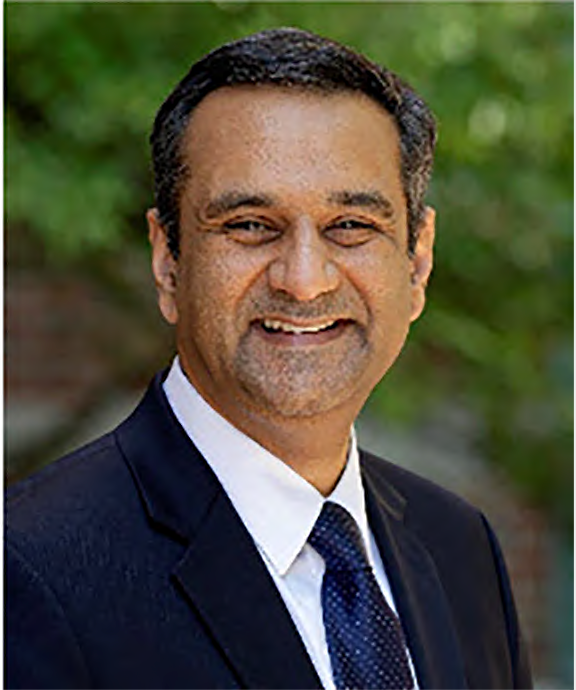
Professor Rohit Bhargava

You and your team focused greatly on studying the spatial constraint that is measured by the shape and structure of tumors and how this affects how individual tumor cells interact with the tumor cellular microenvironment. Particularly, you and your team believed that high-definition Fourier transform infrared (HD-FT-IR) spectroscopic imaging, coupled with machine learning can accurately identify tumor microenvironment components in tissue images. What makes this an important question to solve and what is its significance to medical research and development?
Despite significant advances in cancer biology, we still need to understand cancer progression in its entirety. A greater understanding of its spatial and biochemical composition can lead to new opportunities in diagnostics and therapeutics, for example. We believe that this could be an area where spectroscopic imaging provides a solution. FT-IR imaging offers a full spectral range and, with HD optics, an orderly means to minimize the volume from which high-quality data can be obtained. Machine learning takes this extensive data set and provides insights that are difficult to appreciate manually, while being objective and consistent in assessment. Together, these techniques can help characterize the tumor microenvironment and its influence on cancer progression. There is also a significant opportunity for spectroscopists to drive new technologies, balancing speed, spectral detail, and spatial resolution to provide integrated solutions that can help analyze the complexities of tissue and their microenvironments. In fact, the tissue microenvironment is a general determinant of disease progression and development. We now have a National Institutes of Health (NIH) supported training program in which we educate the next generation at this interface of imaging, computation, and molecular content.
In your article, you and your team talked about a high-definition infrared imaging–based organizational measurement framework (INFORM) that leverages intrinsic chemical contrast of tissue to label unique components of the tumor and its microenvironment. Why is INFORM a great technique for the analysis you are doing?
INFORM is an early-stage tool to show that leveraging the microenvironment information through IR imaging can add value to our ability to distinguish between potentially more and less aggressive colon cancer. The ability to predict the potential lethality of the disease at the time of initial diagnoses can potentially allow us to intervene quickly in those with the greatest need, discover new factors that can help predictions and guide further research. Now that we have this technological demonstration, we can build on this, refine the technique, and translate it to test on patient populations. I should also point out that this study is just the latest example of using the microenvironment to study disease progression. We and other groups have previously also reported association of microenvironmental changes with disease progression. The need now is to further develop technology and explore its use in specific problems, which this study attempted to do.
In your study on predicting tumor behavior, what did you do in this study that is different from what other researchers have done?
The major difference between our work and that of others is that we have championed imaging a diversity of tissue samples, using a tissue microarray format, now with HD FT-IR imaging. This is a time-consuming and onerous task but essential to motivate the development of faster imaging techniques. To our knowledge, this is also the most extensive study (in terms of number of patients) reported thus far with HD-IR imaging. Next, we developed a computational pipeline to utilize this data and explicitly derive parameters from different cell types of the tumor microenvironment. Finally, we translated the development to surgical samples and demonstrated predictive capability from parts of the tumor. Thus, the work establishes a technological pathway from laboratory to clinical samples.
Will varying types of tumor tissue prove to be more difficult to discriminate?
Yes, indeed. The more normal a tumor is, both biochemically and morphologically, the tougher it will be for any technique. However, given the vast potential of spatial-spectral IR data, I think the question we should be asking is: How accurately and optimally can we recognize disease for almost all cancers?
What challenges did you face in developing and implementing microenvironment-based imaging?
The main challenges are the complexity of the microenvironment and a lack of a priori information on which components can be measured in IR with sufficient accuracy to allow us to use them for predictions. These analytical considerations are confounded by the heterogeneity and variation of the disease itself.
Is HD-FT-IR being used for patient treatment at this time? What are the barriers to fully implementing this technique in medical practice?
No FT-IR imaging technique is being used for patient treatment at this time. That is a somewhat premature step for this field. After many years of fundamental progress, we have contributions by many groups to different types of instruments, novel analysis techniques, and demonstrations of various applications. Together, these three areas come together to provide useful solutions that then need to be validated in large patient cohorts. I should caution that, while the data quality of HD-IR is high, high imaging quality (often at the expense of time or signal to noise ratio) is not an automatic technological solution for every problem. A careful matching of the needs, technology development, and validation is needed for a few use cases. This remains the main barrier to further, translational development.
You received the Ellis R. Lippincott Award because of your contributions to the fundamental physics and instrument engineering of mid-IR microscopy and its applications to medical imaging. What are your next steps in furthering your research and what do you think is the future of medical imaging?
I am deeply honored to have received this recognition. It is a testament to the growing field of IR imaging that the work of my research group has been recognized. The last 10 years have seen an accelerating trend of progress with new theoretical understanding, the availability of quantum cascade lasers, many new instrument configurations, and application of IR imaging to a much broader range of problems. Our research continues to push some of these directions as well as engage a broad group of practitioners. We are planning to focus on the development of fast imaging systems, with expanded information content arising from higher quality spatial and spectral data and using it to study the microenvironment in a variety of cancers. We believe that this technology can contribute to medical research and practice by offering a new source of information that is particularly suited to the examination of complex tissues.
Rohit Bhargava is a Founder Professor of Engineering and serves as the Director of the Cancer Center at Illinois of the University of Illinois at Urbana-Champaign. Bhargava graduated with a dual-degree B.Tech. (1996) from the Indian Institute of Technology in New Delhi, India, and received a doctoral degree from Case Western Reserve University in Cleveland, Ohio, in 2000. After a stint at the National Institutes of Health, he has been at the University of Illinois as an Assistant (2005–2011), Associate (2011–2012) and Full (2012–) professor. Bhargava is widely recognized for his research on chemical imaging and advances in theory, instrumentation, and applications in cancer pathology. His innovative teaching and mentoring have also been consistently recognized by the success of his students. He directs the NIH T32-supported Tissue Microenvironment training program and founded the cancer scholars program. In addition to his research and educational activities, Bhargava has served to connect the research community in new and exciting ways to address basic science and engineering questions that surround cancer. He proposed and has served to develop the Cancer Center at Illinois—a basic science center at the convergence of science, engineering, and oncology. Direct correspondence to: rxb@illinois.edu.

New Study Reveals Insights into Phenol’s Behavior in Ice
April 16th 2025A new study published in Spectrochimica Acta Part A by Dominik Heger and colleagues at Masaryk University reveals that phenol's photophysical properties change significantly when frozen, potentially enabling its breakdown by sunlight in icy environments.
AI Shakes Up Spectroscopy as New Tools Reveal the Secret Life of Molecules
April 14th 2025A leading-edge review led by researchers at Oak Ridge National Laboratory and MIT explores how artificial intelligence is revolutionizing the study of molecular vibrations and phonon dynamics. From infrared and Raman spectroscopy to neutron and X-ray scattering, AI is transforming how scientists interpret vibrational spectra and predict material behaviors.