Recording the Raman Spectrum of a Single Molecule
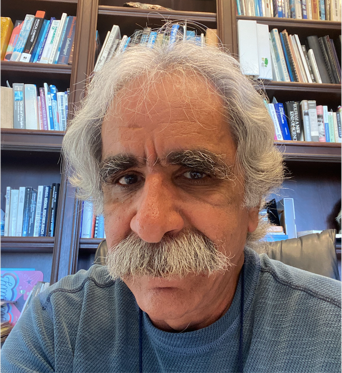
Professor Vartkess Ara Apkarian

Analytical chemists are continually striving to advance techniques to make it possible to observe and measure matter and processes at smaller and smaller scales. Professor Vartkess Ara Apkarian and his team at the University of California, Irvine have made a significant breakthrough in this quest: They have recorded the Raman spectrum of a single azobenzene thiol molecule. The approach, which breaks common tenets about surface-enhanced Raman scattering/spectroscopy (SERS) and tip-enhanced Raman spectroscopy (TERS), involved imaging an isolated azobenzene thiol molecule on an atomically flat gold surface, then picking it up and recording its Raman spectrum using an electrochemically etched silver tip, in an ultrahigh vacuum cryogenic scanning tunneling microscope. For the resulting paper detailing the effort [1], Apkarian and his associates are the 2021 recipients of the William F. Meggers Award, given annually by the Society for Applied Spectroscopy to the authors of the outstanding paper appearing in the journal Applied Spectroscopy. We spoke to Apkarian about this research, and what being awarded this honor means to him and his team. This interview is part of an ongoing series with the winners of awards that are presented at the annual SciX conference. The award will be presented to Apkarian at this fall’s event, which will be held in person in Providence, Rhode Island, September 28–October 1.
What is the significance to the scientific community of obtaining a Raman spectrum from a single molecule?
Improvement of spatial resolution has driven advances in optical microscopy, which has a history of over 1000 years. As far as molecular matter is concerned, seeing its smallest constituent, namely atoms, would be the ultimate limit. We have demonstrated this experimentally for the first time, by showing optical microscopy with a spatial resolution of 1.6 Å, equal to the radius of a silver atom (2). Beyond seeing single molecules, it is possible to wire molecules to photons (3), to use single molecules as complete opto-electronic devices (4), and, more generally, to access the sub-nano frontier in science.
Were there any previous efforts that you found especially useful in advancing this work further, be it a particular scientific approach or merely motivation or inspiration?
Surface-enhanced Raman scattering/spectroscopy (SERS) was discovered in 1974 by noting that the Raman intensity of molecules adsorbed on a roughened silver electrode were ~1 million times stronger than anticipated. Despite the very large literature on principles and applications of SERS, the subject remains a source of fascination, and we are only now reaching a rigorous understanding because of experimental advances. Perhaps the most incisive of these advances is SERS combined with scanning tunneling microscopy (STM) carried out under ultrahigh vacuum (UHV) conditions and cryogenic temperatures (5 K), exemplified by the current paper (1). This was the first direct demonstration that the spectrum of a single molecule can be observed on roughened silver by STM imaging of the molecule, and then picking it up by the tip and recording its spectrum. The observations, which were rigorously analyzed, went against many tenets as stated in the last line of the paper’s abstract: "Tips decorated with asperities break the rules and give unique insights on Raman driven by cavity modes of a plasmonic junction".
Our motivation and inspiration were the years of indirect experimental observations that had generated significant folklore about the subject. Decisive experimental observations were necessary to break through. It took us ~10 years to build the instrument and reach the point where we could carry out the definitive measurements. There are now a handful of laboratories in the world that have built up the same capabilities, such as Zenchao Dong's group in USTC and Martin Wolf's group at the Max Planck in Berlin, and Nan Jiang’s group at University of Illinois, Chicago and the number is growing as the tools become more accessible.
What motivated you to focus on this specific approach to this challenging problem?
The concept that light can be super-focused on a plasmonic wedge (5) or cone was well-established theoretically (6), and the approach of tip-enhanced Raman had already been taken by several groups starting circa 2000. The definitive experiments required atomic control of tip morphology, sub-Å resolution in scanning, and Å-scale stability in the substrate, which could be achieved by combining precision optics and a UHV cryogenic scanning tunneling microscope. This required significant instrumental development, and our earlier reports of atom-resolved optical imaging involved photo-tunneling (7) and electroluminescence with submolecular resolution (8).
What was your process to devise a method to measure the spectrum of a single molecule?
The combination of optics with high collection efficiency and ultrahigh vacuum scanning tunneling microscope operating at 5 K under pristine conditions was necessary to learn the governing physics, and to be able to definitively image a single molecule and record its Raman spectrum.
How was your approach different from what others have previously tried?
Precision instruments were necessary to learn that general notions of tip-enhanced Raman scattering (TERS) were misguided. We developed TERS in the atomistic near-field, by recognizing that on atomically sharp silver needles, the photon converts to charge.
Do you think that your method may be broadly used in laboratories, or will it be a specialized research project for some time to come?
I expect it to be broadly used, and already many new efforts are starting around the world.
Any word whether your findings were incorporated in other efforts? What sort of feedback did you receive?
I find it somewhat ironic that previously the work was rejected twice. One criterion used to that end was that the described work could not be duplicated elsewhere. The body of the work carried by the team is now being recognized and adapted, as evident by the citations of the subsequent works (2-4).
Were there any particularly difficult challenges you encountered in your research?
The effort, which involved significant instrument development and the creation of a dedicated laboratory, required significant marshaling of resources and a long-time commitment, was only possible through Center type support provided by the National science Foundation (NSF). We first proposed the work to NSF in 2007, and created the Center for Chemistry at the Space-Time limit (CaSTL) for the expressed purpose of observing molecular motions with atomistic resolution. The Center had a 10-year lifespan, and delivered on its lofty promise of creating the Chemists' microscope.
Can you elaborate on the strengths that each member of the team bought to the project?
Joonhee Lee has been the team leader (project scientist) from inception to sunset of the CaSTL Center. He has led all aspects of the work, from construction of the instrument to execution of experiments to theoretical analysis, and now has started his own group at the University of Nevada to continue along the same general line of research. Nick Tallarida and Laura Rios were graduate students at the time. They were engaged in the execution of the very intensive experimental work and modeling, both in this project and several other joint publications on related work. The Mars mission is in Nick's good hands who is now at NASA, Jet Propulsion Laboratory (JPL), and the education of next generation scientists is Laura's good hands, as she is now on the faculty at the California Polytechnic Institute.
What are the next steps regarding this research?
This work has already led to a significant body of developments reflected in (2,3,4) and a series of papers in the making, addressing the sub-nano scale in microscopy, molecular devices, molecular electronics, with the key development being the confinement of the photon. Just like the confinement of the electron, which seeded nanoscience, the atomically confined photon (attained at the apex of an atomically sharp STM tip) has opened up the frontier of picoscience, another order of magnitude in miniaturization of devices.
What does being awarded the William F. Meggers award mean to you?
A tremendous amount of dedication and effort was expended by all team members in carrying out the work that appears on several pages of the journal. The cryo-UHV-STM-SERS/TERS work is complex and demanding. The experiments usually demand 24/7 operation over weeks on end. It is therefore satisfying to see an appreciation of the fruit of that labor by the Meggers Award.
References
(1) J. Lee, N. Tallarida, L. Rios, and V.A. Apkarian, Appl. Spectrosc.74(11) 1414 (2020).
(2) J. Lee, K.T. Crampton, N. Tallarida, and V. Ara Apkarian, Nature 568, 7750 (2019).
(3) K.T. Crampton, J. Lee, and V.A. Apkarian, ACS Nano 13, 6363 (2019).
(4) J. Lee, N. Tallarida, X. Chen, L. Jensen, and V. A. Apkarian, Science Advances 4, eaat5472 (2018).
(5) Kh. V. Nerkararyan, Phys. Lett. A 237, 103 (1997).
(6) A. J. Babadjanyan, M. L. Margaryan, and Kh. V. Nerkararyan, J. Appl. Phys. 87, 3785 (2000).
(7) J. Lee, S. Perdue, D. Whitmore, and V. A. Apkarian, J. Chem. Phys.133, 104706 (2010).
(8) J. Lee, S. M. Perdue, A. Rodriguez Perez, and V. A. Apkarian, ACS Nano 8, 54 (2014).

AI-Powered SERS Spectroscopy Breakthrough Boosts Safety of Medicinal Food Products
April 16th 2025A new deep learning-enhanced spectroscopic platform—SERSome—developed by researchers in China and Finland, identifies medicinal and edible homologs (MEHs) with 98% accuracy. This innovation could revolutionize safety and quality control in the growing MEH market.
New Raman Spectroscopy Method Enhances Real-Time Monitoring Across Fermentation Processes
April 15th 2025Researchers at Delft University of Technology have developed a novel method using single compound spectra to enhance the transferability and accuracy of Raman spectroscopy models for real-time fermentation monitoring.
Nanometer-Scale Studies Using Tip Enhanced Raman Spectroscopy
February 8th 2013Volker Deckert, the winner of the 2013 Charles Mann Award, is advancing the use of tip enhanced Raman spectroscopy (TERS) to push the lateral resolution of vibrational spectroscopy well below the Abbe limit, to achieve single-molecule sensitivity. Because the tip can be moved with sub-nanometer precision, structural information with unmatched spatial resolution can be achieved without the need of specific labels.