True Surface Microscopy
Confocal Raman imaging opened the door for many applications in Raman spectroscopy and imaging that were previously unavailable for measurement with conventional (non-confocal) Raman methods. However, high confocality always results in a high focus sensitivity and this can make measurements difficult with rough or inclined samples.
Confocal Raman imaging opened the door for many applications in Raman spectroscopy and imaging that were previously unavailable for measurement with conventional (non-confocal) Raman methods. However, high confocality always results in a high focus sensitivity and this can make measurements difficult with rough or inclined samples. The new, patent pending, True Surface imaging extension resolves these problems and extends Raman imaging to large scale (> 1 × 1 mm2 ) samples without extensive tilt alignment or sample preparation and without comprising the advantages of confocal imaging.
Confocal Raman Imaging
By integrating a Raman spectrometer within a state-of-the-art confocal microscope setup, Raman imaging with a spatial resolution down to 200 nm laterally and 500 nm vertically can be achieved using visible light excitation. Only light from the image focal plane can reach the detector, which strongly increases image contrast and slightly increases resolution. Special filters are used to suppress the reflected laser light while enabling the Raman scattered light to be detected with a spectrometer/CCD camera combination. With this setup, a complete Raman spectrum is acquired at each image pixel, typically taking between 0.7 and 100 ms. The individual spectra are combined to form Raman images consisting of tens of thousands of spectra. From this multispectrum file, an image is generated by integrating over a certain Raman line in all spectra or by evaluating the various peak properties such as peak-width, min/max analysis or peak position. Due to the confocal arrangement, even depth profiling and 3-D imaging are possible if the sample is transparent.
True Surface Microscopy for Topographic Raman Imaging
WITec's exclusive new True Surface Microscopy option makes it possible to perform confocal imaging measurements parallel with and guided by large area topographic scans (> 1 × 1 mm2 ). To achieve this unique capability, the WITec alpha500 microscope series can be equipped with a highly precise sensor for optical profilometry. The large area topographic coordinates from the profilometer measurement are used to perfectly trace the samples surface in either confocal or confocal Raman imaging mode. Sample preparation is reduced to a minimum without having to compromise the confocality of the system. The True Surface Microscopy option is beneficial for many applications, including the characterization of micromechanical, medical, or semiconductor devices, the mapping of functionalized surfaces, or the imaging of bio-medical or pharmaceutical material surface properties. Figure 1a shows a height profile of a pharmaceutical tablet (aspirin) and Figure 1b the same profile with the confocal Raman measurement overlaid. The drugs are labeled red and blue respectively, while the excipient is shown in green (Figure 1c). Topographic variation was more than 300 µm.


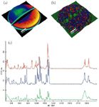
Figure 1a: Topography of an aspirin tablet as obtained with an optical profilometer. Scan range is 12 mm à 12 mm and topographic variation is more than 300 µm. Figure 1b: True Surface Raman image as obtained by using the topography information from Figure 1a with chemical information overlaid. Red and blue are components of the drug, while green is the excipient (300 à 300 = 90,000 spectra, 66 ms integration time/spectrum). Figure 1c: Raman Spectra of the three components.
WITec Instruments Corp.
200 East Broadway Ave. Suite 30, Maryville, TN 37804
tel. (865) 984-4445, fax (865) 984-4441
Email: info@witec-instruments.com
Website: www.WITec-Instruments.com


AI Shakes Up Spectroscopy as New Tools Reveal the Secret Life of Molecules
April 14th 2025A leading-edge review led by researchers at Oak Ridge National Laboratory and MIT explores how artificial intelligence is revolutionizing the study of molecular vibrations and phonon dynamics. From infrared and Raman spectroscopy to neutron and X-ray scattering, AI is transforming how scientists interpret vibrational spectra and predict material behaviors.


Real-Time Battery Health Tracking Using Fiber-Optic Sensors
April 9th 2025A new study by researchers from Palo Alto Research Center (PARC, a Xerox Company) and LG Chem Power presents a novel method for real-time battery monitoring using embedded fiber-optic sensors. This approach enhances state-of-charge (SOC) and state-of-health (SOH) estimations, potentially improving the efficiency and lifespan of lithium-ion batteries in electric vehicles (xEVs).


New Study Provides Insights into Chiral Smectic Phases
March 31st 2025Researchers from the Institute of Nuclear Physics Polish Academy of Sciences have unveiled new insights into the molecular arrangement of the 7HH6 compound’s smectic phases using X-ray diffraction (XRD) and infrared (IR) spectroscopy.

