Spectroscopy
The application note gives an overview of several pharmaceutical dosage forms and drug systems investigated by topographic confocal Raman imaging which facilitates accurate chemical characterization of even rough and inclined samples.
Spectroscopy
The application note gives an overview of several pharmaceutical dosage forms and drug systems investigated by topographic confocal Raman imaging which facilitates accurate chemical characterization of even rough and inclined samples.
Application Notebook
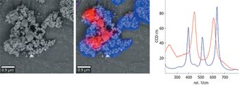
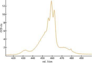
RISE microscopy, the combination of Raman imaging and scanning electron microscopy, is a powerful new analytical method for the analysis and interrelation of the structure and composition of microscopic samples. The integration of both imaging techniques in one instrument avoids shuttling the sample from one microscope to another. Here we demonstrated its usefulness in the identification, discrimination, and localization of polymorphs of titanium dioxide.
Application Notebook
Since Sir C.V. Raman discovered the Raman effect in the 1920s, it has become essential to many fields of research and industry.
Spectroscopy
Confocal Raman microscopy is an excellent tool for true 3D chemical imaging. In this application note the technique for ultrasensitive Raman imaging is highlighted along with various examples of applications.
Spectroscopy
This application note describes how the spectral and spatial resolution of a confocal Raman microscope can be ptimized by the usage of ideal optical and mechanical microscope components.
Spectroscopy
Confocal Raman imaging is a nondestructive, label-free method to study living cells, bacteria, and tissues. It includes new data on biofilms, human skin, vessels and liver tissue.
Spectroscopy
Confocal Raman imaging can be effectively used for forensic investigations such as fingerprint or ink analyses.
Spectroscopy
Raman, AFM and SEM imaging provide information regarding the characterization of chemical and structural properties. In this application note you will gain an overview of the WITec imaging techniques through various application examples from graphene research.
Spectroscopy
This application note introduces topographic confocal Raman Imaging as a flexible, noninvasive and non-destructive tool for the chemical and molecular characterization of archaeological specimens.
Spectroscopy
The application note gives an overview of several pharmaceutical dosage forms and drug systems investigated by topographic confocal Raman imaging which facilitates accurate chemical characterization of even rough and
Application Notebook
RISE microscopy is a novel correlative microscopy technique that combines SEM and confocal Raman imaging. Through RISE microscopy ultra-structural surface properties can be linked to molecular compound information.
Application Notebook
Newly developed software tools provide various functions for the acquisition, evaluation, and processing for high-resolution 3D Raman images.
Application Notebook
Confocal Raman imaging opened the door for many applications in Raman spectroscopy and imaging that were previously unavailable for measurement with conventional (non-confocal) Raman methods.

Application Notebook
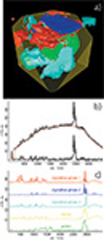
For the characterization of the properties of a sample with Raman spectroscopy, an ultrasensitive confocal Raman microscope allows the acquisition of a Raman image stack revealing 3-D information on the distribution of the chemical compounds.

Application Notebook
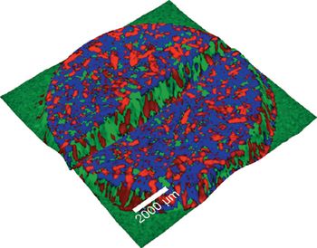
The development of advanced polymeric materials requires detailed information about the phase separation process on the nanometer scale. Confocal Raman microscopy contributes to the analysis of such materials by visualizing the distribution of individual components based on the unique Raman spectra for different polymeric materials. Using a confocal setup, polymer domains can be imaged three-dimensionally with a resolution down to 200 nm. As a Raman image typically consists of tens of thousands of spectra, a powerful data analysis software is essential in order to extract the relevant information. Hidden structures in the images should ideally be visualized automatically, ensuring an objective and consistent interpretation of the imaging data.
Application Notebook