Raman Microanalysis in the Forensic Laboratory: Identification and Characterization of Suspect Solids, Liquids, and Powders
Due to its high information, vibrational content, the Raman spectrum provides important and specific molecular information. In this article, Raman microanalysis and instrumentation is discussed with specific application to the forensic science laboratory, where evidence integrity is of the utmost importance.
Infrared spectroscopy is used extensively for compound identification and routine analysis in the forensic science laboratory. Raman spectroscopy is an attractive technique that supplies complementary vibrational information to infrared. Applying both infrared and Raman spectroscopy to a given forensic problem allows a full vibrational characterization to be obtained that can reveal new and important facts about the problem under investigation.
Raman spectrometers have benefited from years of steady improvement in instrument design, which have led to instrumentation that is compact, incorporates extensive automation, and is fully validatable to strict regulatory standards. In contrast to Raman spectrometers of the past, modern Raman spectrometers are simple to operate, easy to maintain, and provide accurate and reproducible answers to complex problems. As a result, there has been renewed interest in employing Raman spectroscopic characterization in the field of a forensic science. In this article, Raman spectroscopy and instrumentation will be presented and discussed with specific application to the forensic science laboratory.
Raman Spectroscopy
Raman spectroscopy is well suited to chemical analysis in forensic science. The Raman spectrum provides molecular information that is unique to the molecules present, allowing Raman to be used as a method for molecular identification. Raman measurements are fast, with acquisition times typically requiring only a few seconds. The analysis is a noncontact, nondestructive measurement. Raman measurements are insensitive to water and consequently characterization of aqueous samples difficult to obtain with other techniques is obtained easily using Raman spectroscopy. There is little or no sample preparation required, making sampling extremely flexible. Measurements can be performed on microscope slides or directly through glass bottles or through vials. In the forensic science laboratory, integrity of the chain of evidence is very important and measures must be taken to insure samples remain free of contamination after evidence is collected at the scene of the crime. With Raman spectroscopy, chain of evidence concerns can be alleviated by sampling directly through plastic evidence bags.
Raman spectroscopy has long been recognized as a valued technique in research. Important progress has been made in instrument design, which has improved the performance of Raman instrumentation. Development most recently has been focused on incorporating features that make instrumentation more practical. While the practice of Raman spectroscopy used to be the realm of highly specialized, trained experts, today's most advanced, modern Raman instruments are integrated completely into a single unit, which is computer-controlled, interlocked for laser safety, and with automated protocols for calibration and alignment. These instruments are truly walk-up instruments that require only a minimal training for the user to be able to acquire accurate and reproducible measurements. These advances in instrument design have eliminated many if not all the barriers to the implementation of Raman spectroscopy as a powerful tool alongside the existing techniques employed in the forensic laboratory.
There are two different types of Raman spectrometers; visible dispersive Raman and Fourier-transform Raman (FT-Raman). Dispersive Raman instruments generally are more sensitive, and are well-suited for small samples and mapping applications where high spatial resolution is required. FT-Raman spectrometers provide fast and accurate results with the benefit of fluorescence-free Raman spectra. The Raman spectra presented here were acquired using a Thermo Electron Corporation Nicolet Almega dispersive Raman microscope and a Thermo Electron Corporation FT-Raman module. Computer control, data acquisition, and data processing for both instruments were achieved through Thermo Electron's OMNIC suite of software.
Raman spectroscopy is ideal for a number of analyses in the forensic laboratory. These include but are not limited to: illicit drug identification, fiber characterization, trace analysis of explosive materials, and paint chip analysis. Several examples are presented here.
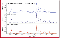
Figure 1. (top) The dispersive Raman, (middle) FT-Raman, and (bottom) Raman library reference spectra of the controlled substance g-butyrolactone (GBL), a precursor to GBH (Liquid Ecstasy).
Illicit Drug Analysis
Raman spectroscopy offers safe and rapid analysis of many illicit drugs. In many cases the submitted materials can be analyzed directly through glass vials or plastic evidence bags, thereby avoiding potential evidence tampering. Figure 1 shows the Raman spectrum of γ-butyrolactone (GBL). GBL is a controlled substance that is a precursor to GHB (known as Liquid Ecstasy or Easy Lay). It is a so-called "rave" drug and a major problem drug. It is a liquid and is analyzed easily using FT or dispersive Raman. Comparison of the collected spectra with established spectral libraries leads to definitive compound identification. As a further example, Figure 2 presents the Raman spectrum of another commonly encountered illicit street drug: cocaine HCl.
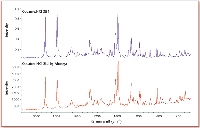
Figure 2. (top) The dispersive Raman spectrum of cocaine HCl and (bottom) reference cocaine HCl.
Fiber Identification
Fibers found at crime scenes can be of prime importance to some forensic investigations. Dispersive Raman microscopy is particularly well-suited to trace evidence characterization. The technique again requires no sample preparation. The dispersive Raman spectra for three different fibers are shown in Figure 3. In each case, fibers were placed on a glass microscope slide and the Raman spectra were collected through the attached Raman microscope using a 10x objective. The different fibers are rapidly and easily distinguished from each other, and can be identified using spectral library searches.
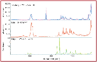
Figure 3. The FT-Raman spectra of three fibers. A spectral library search using the spectral database in the OMNIC software readily identified the fibers.
Paint Chip Characterization
Law enforcement is called upon routinely to investigate automobile accidents. In "hit and run" accidents, only traces of evidence remain at the accident scene. Important clues to the identity of the vehicle involved in the accident can reside in a small paint chip. Paint chips typically contain multiple layers (for example, primer, bottom coat, and so forth) each with a distinct chemical composition. Raman microanalysis of the paint chip layers will aid in identifying the vehicle involved in the crime. Figure 4 shows a cross-sectional view of a paint chip subjected to Raman microanalysis. No sample preparation was involved, other than placing the paint chip in a clay mount. Raman microanalysis characterizes each of the layers in the paint chip by simply moving the stage of the microscope to each of the targeted areas of interest. Figure 4 shows the distinct Raman spectra that are obtained from each layer in the paint chip.
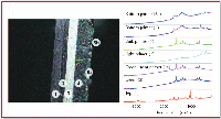
Figure 4. (left) Cross-sectional view of a paint chip with corresponding dispersive Raman spectra.
Conclusion
Raman spectroscopy is now recognized to be an indispensable tool for the forensic laboratory. It has a number of inherent advantages over other techniques. Major advances in instrumentation have led to greater reliability, reproducibility, a much higher level of automation, and greater ease of use. Instruments now are much easier to maintain than the instruments that were available as recently as five years ago. Raman spectroscopy is a powerful technique that can provide rapid answers to some of the most complex problems encountered in the forensic laboratory.
Mark H. Wall is a product specialist, Raman products, for Thermo Electron Corp., Molecular Spectroscopy Div. (Madison WI). E-mail: mark.wall@thermo.com. Linda De Noble and Richard Hartman are with the New Hampshire Department of Safety, State Police Forensic Laboratory.
High-Speed Laser MS for Precise, Prep-Free Environmental Particle Tracking
April 21st 2025Scientists at Oak Ridge National Laboratory have demonstrated that a fast, laser-based mass spectrometry method—LA-ICP-TOF-MS—can accurately detect and identify airborne environmental particles, including toxic metal particles like ruthenium, without the need for complex sample preparation. The work offers a breakthrough in rapid, high-resolution analysis of environmental pollutants.
New Study Reveals Insights into Phenol’s Behavior in Ice
April 16th 2025A new study published in Spectrochimica Acta Part A by Dominik Heger and colleagues at Masaryk University reveals that phenol's photophysical properties change significantly when frozen, potentially enabling its breakdown by sunlight in icy environments.
AI-Driven Raman Spectroscopy Paves the Way for Precision Cancer Immunotherapy
April 15th 2025Researchers are using AI-enabled Raman spectroscopy to enhance the development, administration, and response prediction of cancer immunotherapies. This innovative, label-free method provides detailed insights into tumor-immune microenvironments, aiming to optimize personalized immunotherapy and other treatment strategies and improve patient outcomes.