Organic Nitrogen Compounds VIII: Imides
Spectroscopy
Here, we continue our examination of the infrared (IR) spectra of organic nitrogen compounds with imides, which are a common chemical intermediate. IR can be used not only to identify imides, but also to distinguish between straight chain and cyclic imides. We explain how.
We continue our examination of the infrared spectra of organic nitrogen compounds with imides. Imides are a common chemical intermediate, and the engineering polymer Kapton is a polyimide. These molecules have an unusual structure consisting of two carbonyl groups connected by a nitrogen atom. Their spectra are unique as well since some imides exhibit two C=O stretching peaks. Infrared spectroscopy can be used not only to identify imides, but distinguish between straight chain and cyclic imides as well.
We are on installment eight of our series on the infrared spectra of organic nitrogen compounds. Who would have thought there were so many of them? The current installment looks at the spectra of imides. These molecules are not to be confused with amides, which we studied in the last two installments (1,2), even though their names and structures are similar. Amides have many pronunciations (2), my preferred one being ay-mides. For imides the pronunciation is im-id, where the first syllable rhymes with the “im” in “immobile,” and the last syllable rhymes with the word “id,” which Freud told us is a part of the human psyche (3).
The structural framework of an imide is seen in Figure 1.
Figure 1: The structural framework of the imide functional group.
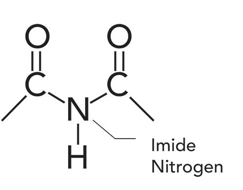
Recall that amides (1,2) have a carbonyl group attached to a nitrogen. An imide, however, is like an amide on steroids, as it has two carbonyl groups connected by a central nitrogen atom, as seen in Figure 1. As we will see, the spectra of amides and imides have similarities. Imides also have some similarity to acid anhydrides. Recall (4) that an acid anhydride is two carbonyl groups connected by a central oxygen atom. The double carbonyl group in acid anhydrides gives rise to a double carbonyl stretch. Some imides exhibit this type of spectral feature as well.
Note that the central nitrogen atom in the imide functional group is called the imide nitrogen. This nitrogen atom may have a hydrogen atom bonded to it, as shown in Figure 1, or have a carbon bearing substituent such as an alkyl chain or phenyl group attached. In either case, the molecule is still classified as an imide. Imides come in two varieties; straight chain and cyclic. Below, we will discuss how to use infrared spectroscopy to distinguish these two imide types from each other.
The Infrared Spectroscopy of Imides
The infrared spectrum of a cyclic imide, phthalimide, is seen in Figure 2. Note that this imide has a hydrogen attached to the imide nitrogen. Imides like this will exhibit an imide N-H stretch, which falls at 3200±50 cm-1 (going forward, all peak positions will be in cm-1, even if not noted). This peak is labeled A in Figure 2. The N-H stretching peak position is the same for both cyclic and straight chain imides.
Figure 2: The infrared spectrum of phthalimide, (C8H5NO2), a cyclic imide.
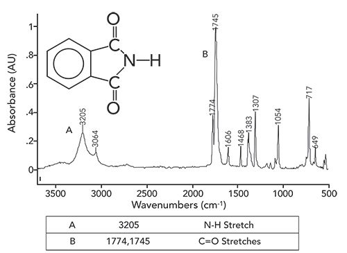
Note that, like the N-H stretches of amines (5) and amides (1,2), this peak is of medium width, medium intensity, and shows up in the same region as O-H stretches. However, it is easy to distinguish OH from NH stretches since the former are much more intense and broader than the latter. Of course, if there is a carbon atom attached to the imide nitrogen, there will be no N-H bond, and hence no N-H stretching peak. Thus, unfortunately not all imides have an N-H stretching peak.
One of the spectral signatures of a cyclic imide is a double carbonyl stretch. These peaks are labeled B in Figure 2, and fall at 1774 and 1745. In general, these two peaks are found from 1790 to 1735, and 1750 to 1680 for cyclic imides. Acid anhydrides also have a double carbonyl stretch that falls in this range (4). However, acid anhydrides do not contain nitrogen, and hence will not exhibit an N-H stretch. Additionally, acid anhydrides have a strong C-O stretching peak that is missing from imides, because they do not contain this kind of bond. Thus, the diagnostic pattern for a cyclic imide is a single N-H stretch (when present) in combination with a double carbonyl stretch.
Note from the structure of phthalimide that all its carbons are unsaturated. Recall (6) that such molecules only have C-H stretches above 3000. This is the case in Figure 2, where the aromatic C-H stretching peak is at 3064, and there are no C-H stretching peaks below 3000. This C-H stretching peak appears as a shoulder on the broader N-H stretching envelope.
Unlike cyclic imides, straight chain imides have only one C=O stretch that falls from 1740-1670. This means cyclic and straight chain imides can be distinguished from each other, since cyclic imides have two carbonyl stretches, whereas straight chain imides have one C=O stretching peak. However, the N-H and C=O stretching peaks of straight chain imides fall in the same range as secondary amides (1). This means it may be difficult to distinguish between a straight imide and secondary amide based on their infrared spectra by themselves. In situations such as this, other measurements, such as physical properties, mass spectra, NMR spectra, or Raman spectra may be needed to get the job done. This is nothing to be alarmed at. As I have been mentioning throughout this column series (7), determining the complete structure of a molecule solely from its infrared spectrum is not always possible. Other data are sometimes required, and that is why chemists have developed multiple instrumental techniques to determine molecular structure. In my experience, most of the industrially important imides are cyclic, and you will run across this type of imide more often than straight chain imides. A summary of the imide group wavenumbers is seen in Table I.

Conclusions
The imide functional group consists of a central nitrogen atom, the imide nitrogen, attached to two carbonyl groups. Imides come in two varieties; cyclic and straight chain. The two can be distinguished from each other because cyclic imides have two carbonyl stretches, whereas straight chain imides have one. The diagnostic peak pattern for cyclic imides is a double carbonyl stretch combined with an N-H stretch (when present). Unfortunately, the infrared spectra of straight chain imides are similar to those of secondary amides, making it sometimes difficult to distinguish between the two based on their infrared spectra alone. Other information may be needed to differentiate between them.
References
- B.C. Smith, Spectroscopy 35(1), 10–15 (2020).
- B.C. Smith, Spectroscopy 34(11), 30–33 (2019).
- https://en.wikipedia.org/wiki/Id,_ego_and_super-ego#Id
- B.C. Smith, Spectroscopy 33(3), 16–20 (2018).
- B.C. Smith, Spectroscopy 34(3), 22–25 (2019).
- B.C. Smith, Spectroscopy 31(11), 28–34 (2016).
- B.C. Smith, Spectroscopy 30(1), 16–23 (2015).
- B.C. Smith, Spectroscopy 34(1), 10–15 (2019).
- B.C. Smith, Spectroscopy 32(9), 31–36 (2017).
- B.C. Smith, Spectroscopy 31(5), 36–39 (2016).

Brian C. Smith, PhD, is the founder and CEO of Big Sur Scientific, a maker of portable mid-infrared cannabis analyzers. He has over 30 years of experience as an industrial infrared spectroscopist, has published numerous peer reviewed papers, and has written three books on spectroscopy. As a trainer, he has helped thousands of people around the world improve their infrared analyses. In addition to writing for Spectroscopy, Dr. Smith writes a regular column for its sister publication Cannabis Science and Technology and sits on its editorial board. He earned his PhD in physical chemistry from Dartmouth College. He can be reached at: SpectroscopyEdit@MMHGroup.com

AI Shakes Up Spectroscopy as New Tools Reveal the Secret Life of Molecules
April 14th 2025A leading-edge review led by researchers at Oak Ridge National Laboratory and MIT explores how artificial intelligence is revolutionizing the study of molecular vibrations and phonon dynamics. From infrared and Raman spectroscopy to neutron and X-ray scattering, AI is transforming how scientists interpret vibrational spectra and predict material behaviors.
Real-Time Battery Health Tracking Using Fiber-Optic Sensors
April 9th 2025A new study by researchers from Palo Alto Research Center (PARC, a Xerox Company) and LG Chem Power presents a novel method for real-time battery monitoring using embedded fiber-optic sensors. This approach enhances state-of-charge (SOC) and state-of-health (SOH) estimations, potentially improving the efficiency and lifespan of lithium-ion batteries in electric vehicles (xEVs).
New Study Provides Insights into Chiral Smectic Phases
March 31st 2025Researchers from the Institute of Nuclear Physics Polish Academy of Sciences have unveiled new insights into the molecular arrangement of the 7HH6 compound’s smectic phases using X-ray diffraction (XRD) and infrared (IR) spectroscopy.